logo
AAT Bioquest

Screen Quest™ Colorimetric Glucose Uptake Assay Kit

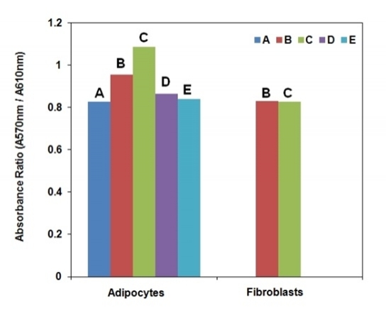
Glucose transport systems are responsible for transporting glucose across cell membranes. Measuring uptake of 2-deoxyglucose (2-DG), a glucose analog, in tissues and cells is widely accepted as a reliable method to estimate the amount of glucose uptake and to investigate the regulation of glucose metabolism and mechanism of insulin resistance. The 2-DG uptake is commonly determined by using non-metabolized 2-DG labeled with tritium or C14. However, routine use of a radiolabelled probe is costly and requires a tedious special handling procedure. AAT Bioquest's Screen Quest™ Colorimetric Glucose Uptake Assay Kit provides a sensitive and non-radioactive assay in tissues or cultured cells. In this assay 2-DG is taken up by glucose transporters, and metabolized to 2-DG-6-phosphate (2-DG6P). The non-metabolizable 2-DG6P accumulates in the cells, and is proportional to glucose uptake by cells. The accumulated 2-DG6P is enzymatically oxidized and generates NADPH, which is specifically monitored by a chromogenic NADPH sensor. The signal can be read by a absorption microplate reader by reading the OD ratio at wavelength 570 nm to 610 nm.

Example protocol

AT A GLANCE

Protocol summary

  1. Plate cells and treat the cells as desired
  2. Add 10 µL/well 2-DG and incubate at 37oC for 20-40 minutes
  3. Wash cells and lyse cells
  4. Add 50 µL/well of 2-DG Uptake Assay working solution
  5. Incubate at RT for 30 to 120 minutes
  6. Monitor OD ratio increase at 570/610 nm

Important notes
Thaw all the kit components at room temperature before starting the experiment. The above mentioned protocol is for one 96-well plate.

PREPARATION OF STOCK SOLUTION

Unless otherwise noted, all unused stock solutions should be divided into single-use aliquots and stored at -20 °C after preparation. Avoid repeated freeze-thaw cycles.

KRPH Buffer stock solution (1X):
Add 20 mL of KRPH Buffer (5X) (Component H) to 80 mL of deionized water and mix them well. Note: 50 mL volume of 1× KRPH Buffer is enough for approximately one 96-well plate. Prepare the needed volume proportionally. Store the unused 1×KRPH at 4oC or -20oC.

PREPARATION OF WORKING SOLUTION

1. NADP working solution:
Add 100 µL of H2O into the vial of NADP (Component G) and mix them well.

2. Enzyme Probe working solution:
Add 5 mL of Assay Buffer (Component F) into the bottle of Enzyme Probe (Component E) and mix them well.

3. 2-DG Uptake Assay working solution:
Add 100 µL of NADP working solution into the Enzyme Probe working solution and mix them well. Note: These quantities are good for one 96-well plate.

SAMPLE EXPERIMENTAL PROTOCOL

Important: This protocol can be used as guidelines to culture 3T3-L1 adipocytes for 2-DG uptake.

  1. Plate cells in growth medium at 50,000-80,000 cells/well/100 µL/96-well or 12,500-20,000 cells/well/25 µL/384-well black wall/clear bottom cell culture Poly-D lysine plate for 4-6 hours before experiment.

  2. Remove the cell plate from the incubator, aspirate the medium from the wells, and deprive the cells with 100 µL/well (96 well-plate) or 25 µL/well (384 well-plate) serum free medium. Incubate the cells at 37oC, 5% CO2 incubator for 6 hours to overnight.

  3. Remove the cell plate from the incubator, aspirate the medium from the wells, and gently wash the cells twice with 100 µL/well 1× KRPH buffer.

  4. Add 90 µL/well Glucose Uptake Buffer (Component B) and incubate the cells at 37oC, 5% CO2 incubator for 1 hour.

  5. Stimulate with or without insulin or compound of test for 20 min. Add 10 µL/well of the 10× insulin solution to a final concentration of 1 µM or 10× compound solution of test. And also add 10 µL insulin vehicle buffer or compound vehicle buffer to the untreated wells as control, and incubate at 37oC, 5% CO2 incubator for 20 mins.

  6. For glucose uptake inhibition study, add 10× Phloretin to a final concentration of 200 uM or inhibitors of test, and incubate at 37oC, 5% CO2 for 2-5 mins. Note: 10 mL inhibitor vehicle buffer is suggested to be added to both the insulin treated and untreated wells as control. Phloretin treated cells can be used as positive control.

  7. Add 10 µL/well 2-DG solution (Component A) to each well, and incubate at 37oC, 5% CO2 incubator for 20-40 mins. For negative controls, leave some wells untreated with insulin, inhibitor and 2-DG.

  8. After treatment, remove solution in each well and gently wash cells 3 times, 100 µL/well with KRPH Buffer to remove the extra 2-DG from the solution. Remove KRPH Buffer from the wells.

  9. Add 25 µL/well Acidic Lysis Buffer (Component C) to each well and incubate at 37oC for 20 min to lyse the cells. And the 2DG uptake assay mixture could be prepared in the meantime.

  10. Add 25 µL/well Neutralization Buffer (Component D) to each well, mix thoroughly, leave at room temperature for 5-10 minutes to neutralize the cell lysate.

  11. Add 50 µL of 2DG Uptake Assay working solution to each well of 2-DG6P standard (Not provided) or cell lysate.

  12. Incubate the reaction at room temperature for 30 minutes to 2 hours, protected from light.

  13. Monitor the absorbance ratio increase at 570/610 nm with an absorbance plate reader.

Citations

View all 13 citations: Citation Explorer
Revealing the Mechanism of Esculin in Treating Renal Cell Carcinoma Based on Network Pharmacology and Experimental Validation
Authors: Chen, Zixuan and Wang, Cunzhou and Cai, Yuesong and Xu, An and Han, Chengtao and Tong, Yanjun and Cheng, Sheng and Liu, Min
Journal: Biomolecules (2024): 1043
TFAIP6 facilitates hepatocellular carcinoma cell glycolysis through upregulating c-Myc/PKM2 axis
Authors: Duan, Kecai and Fang, Kunpeng and Sui, Chengjun
Journal: Heliyon (2024): e30959
Exosomes from PYCR1 knockdown bone marrow mesenchymal stem inhibits aerobic glycolysis and the growth of bladder cancer cells via regulation of the EGFR/PI3K/AKT pathway
Authors: Li, Zhuo and Jiang, Ying and Liu, Jian and Fu, Huifeng and Yang, Quan and Song, Wei and Li, Yuanwei
Journal: International Journal of Oncology (2023): 1--13
METTL5 stabilizes c-Myc by facilitating USP5 translation to reprogram glucose metabolism and promote hepatocellular carcinoma progression
Authors: Xia, Peng and Zhang, Hao and Lu, Haofeng and Xu, Kequan and Jiang, Xiang and Jiang, Yuke and Gongye, Xiangdong and Chen, Zhang and Liu, Jie and Chen, Xi and others,
Journal: Cancer Communications (2023)

References

View all 115 references: Citation Explorer
Inhibition of the sodium glucose co-transporter-2: its beneficial action and potential combination therapy for type 2 diabetes mellitus
Authors: Chen LH, Leung PS.
Journal: Diabetes Obes Metab. (2013)
Hepatic expression and cellular distribution of the glucose transporter family
Authors: Karim S, Adams DH, Lalor PF.
Journal: World J Gastroenterol (2012): 6771
Regulation of glucose transporter translocation in health and diabetes
Authors: Bogan JS., undefined
Journal: Annu Rev Biochem (2012): 507
New possibility in the oral glucose lowering treatment of type 2 diabetes mellitus: sodium-glucose co-transporter-2 inhibitors
Authors: Simonyi G., undefined
Journal: Orv Hetil (2012): 695
Glucose transporter type I deficiency syndrome: epilepsy phenotypes and outcomes
Authors: Pong AW, Geary BR, Engelstad KM, Natarajan A, Yang H, De Vivo DC.
Journal: Epilepsia (2012): 1503
Page updated on September 18, 2024

Ordering information

Price
Unit size
100 Tests
500 Tests
Catalog Number
Quantity
Add to cart

Additional ordering information

Telephone1-800-990-8053
Fax1-800-609-2943
Emailsales@aatbio.com
InternationalSee distributors
Bulk requestInquire
Custom sizeInquire
Technical SupportContact us
Purchase orderSend to sales@aatbio.com
ShippingStandard overnight for United States, inquire for international
Request quotation

Storage, safety and handling

H-phraseH303, H313, H333
Hazard symbolXN
Intended useResearch Use Only (RUO)
R-phraseR20, R21, R22
UNSPSC12352200

Platform

Absorbance microplate reader

Absorbance570, 610 nm
Recommended plateClear bottom

Components

Measurement of 2DG uptake in differentiated 3T3-L1 adipocytes and 3T3-L1 fibroblasts. Assays were performed with Screen Quest™ Colorimetric Glucose Uptake Assay Kit in a black wall/clear bottom cell culture Poly-D lysine plate using a SpectraMax (Molecular Devices) microplate reader. (A: Negative Control, no insulin no 2-DG treatment. B: 2DG uptake in the absence of insulin. C: 2DG uptake in the presence of 1mM insulin. D: 2DG uptake in the presence of 1mM insulin and 200 mM phloretin. E: 2DG uptake in the presence of insulin 1mM and 5mM D-Glucose.) 
Measurement of 2DG uptake in differentiated 3T3-L1 adipocytes and 3T3-L1 fibroblasts. Assays were performed with Screen Quest™ Colorimetric Glucose Uptake Assay Kit in a black wall/clear bottom cell culture Poly-D lysine plate using a SpectraMax (Molecular Devices) microplate reader. (A: Negative Control, no insulin no 2-DG treatment. B: 2DG uptake in the absence of insulin. C: 2DG uptake in the presence of 1mM insulin. D: 2DG uptake in the presence of 1mM insulin and 200 mM phloretin. E: 2DG uptake in the presence of insulin 1mM and 5mM D-Glucose.)