Screen Quest™ Live Cell cAMP Assay Service Pack
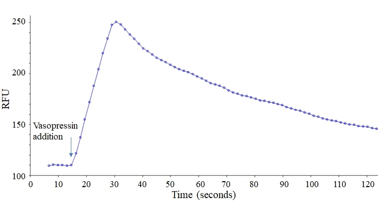
G protein coupled receptors (GPCR) are one of the largest receptor classes targeted by drug discovery programs. Calcium flux (coupled via Gq pathway) assay is a preferred method in drug discovery for screening GPCR targets. However, over 60% of the known GPCRs signal through adenylyl cyclase activity coupled to cAMP. Most of the existing cAMP assays not only require cell lysis but also lack both temporal and spatial resolution. Screen Quest™ Live Cell cAMP Assay Service Pack provides the real-time monitoring of intracellular cAMP change in a high-throughput format without a cell lysis step. The assay works through the cell lines that contain either an exogenous cyclic nucleotide-gated channel (CNGC) or the promiscuous G-protein, Gα16. The channel is activated by elevated levels of intracellular cAMP, resulting in ion flux and cell membrane depolarization which can be detected with either a fluorescent calcium (such as Calbryte 520 AM, Cal-520 AM, Fluo-8 AM, or Fluo-4 AM and corresponding no wash calcium kits) or a fluorescent membrane potential dye. Co-expression of Gα16 with specific non-Gq-coupled receptors will result in the generation of an intracellular calcium signal upon receptor stimulation. The Screen Quest™ Live Cell cAMP Assay Service Pack provides both cell lines and reagents for the measurement of intracellular cAMP changes with a FLIPR, a FDSS or other equivalent fluorescence microplate readers. It has been successfully used to measure Gs and Gi coupled GPCR activity.
Example protocol
AT A GLANCE
Protocol summary
- Prepare cells for transfection
- Prepare Transfectamine™ 5000-DNA mixture
- Add Transfectamine™ 5000-DNA mixture to the cell culture, incubate overnight
- Transfer the transfected cells to a 96-well plate 24-30 hours after transfection, and incubate the culture overnight
- Add Calbryte™ 520 NW dye-loading solution (100 µL/well for a 96-well plate)
- Incubate at room temperature or 37 °C for 30-60 minutes
- Monitor the fluorescence intensity at Ex/Em = 490/525 nm
Important
Thaw the kit components at room temperature before starting the experiment.CELL PREPARATION
- Seed the cells such that they will be ~60-70% confluent at the time of transfection.
- Replace with fresh growth medium before transfection. For example, replace with 2 mL of medium per well for 6-well plates and 6 mL of medium for 10 cm plates.
PREPARATION OF STOCK SOLUTIONS
Unless otherwise noted, all unused stock solutions should be divided into single-use aliquots and stored at -20 °C after preparation. Avoid repeated freeze-thaw cycles.
Note 20 µL of Calbryte™ 520NW stock solution is enough for one plate. Unused Calbryte™ 520NW stock solution can be aliquoted and stored at < -20 °C for more than one month if the tubes are sealed tightly. Protect from light and avoid repeated freeze-thaw cycles.
Calbryte™ 520NW stock solution
Add 20 µL (For Cat# 36382 and 36383), 200 µL (For Cat# 36384) of DMSO (Component F) into the vial of Calbryte™ 520NW (Component C), and mix them well.Note 20 µL of Calbryte™ 520NW stock solution is enough for one plate. Unused Calbryte™ 520NW stock solution can be aliquoted and stored at < -20 °C for more than one month if the tubes are sealed tightly. Protect from light and avoid repeated freeze-thaw cycles.
PREPARATION OF WORKING SOLUTION
1. 1X Assay Buffer
Mix 9 mL of HHBS (Component E) with 1mL of 10X Pluronic® F127 Plus (Component D), and mix them well.2. Calbryte™ 520NW working solution
Add 20 µL of Calbryte™ 520 NW stock solution into 10 mL of 1X Assay Buffer, and mix them well.Note The working solution is stable for at least 2 hours at room temperature.
3. TRANSFECTAMINE™ 5000-DNA MIXTURE
- Add 15 µL of ddH2O to the vial of Ga16 DNA (Component A), mix well to have the final concentration of 1 µg/µL.
Note Skip this step for Cat# 36384. Component A is already provided in solution form and is ready to use. - Mix 3 µg of DNA [for example, 1.5 µg of Ga16 DNA (Component A) and 1.5 µg DNA of the GPCR that you are interested] with 200 µL of serum-free medium.
- Add 9 µL of Transfectamine™ 5000 (Component B) to the mixture from Step 1.
- Mix well and incubate at room temperature for 20 minutes.
Note The ratio of Transfectamine™ 5000 and DNA need to be optimized for different cell lines. In general, the ratio for Transfectamine™ 5000 Transfection Reagent (µL) to DNA (µg) should be 3-5 µL : 1 µg.
Component | 6 well plate (per well) | 10 cm plate |
Fresh culture medium | 2 mL | 6 mL |
Plasmid | ~3 µg | 10~15 µg |
Serum-free medium | 200 µL | 600 µL |
Transfectamine™ 5000 Transfection Reagent | ~9 µL | ~30-45 µL |
SAMPLE EXPERIMENTAL PROTOCOL
Transfection and Translocation protocol
- Add Transfectamine™ 5000 -DNA mixture to the culture plate and incubate overnight.
Note The recombinant protein can start to be detected as early as 16 hours after transfection. The maximal expression level may be observed 72~96 hours after transfection. - Transfer the transfected cells to a 96-well plate 24-30 hours post transfection and incubate overnight.
Note For adherent cells: Plate cells overnight in the growth medium at 40,000 to 80,000 cells/well/100 µL for a 96-well plate.
Note For non-adherent cells: Centrifuge the cells from the culture medium and then suspend the cell pellet in cell growth medium or HHBS at 125,000 to 250,000 cells/well/100 µL for a 96-well poly-D lysine plate. Centrifuge the plate at 800 rpm for 2 minutes with brake off prior to the experiments.
Note Each cell line should be evaluated on the individual basis to determine the optimal cell density for the intracellular calcium mobilization.
Run calcium assay
- Add 100 µL/well (96-well plate) of Calbryte™ 520NW working solution into the cell plate.
- Incubate the dye-loading plate in a cell incubator for 30 minutes, and then incubate the plate at room temperature for another 15-30 minutes.
Note If the assay requires 37 °C, perform the experiment immediately without further room temperature incubation.
Note If the cells can function well at room temperature for longer time, incubate the cell plate at room temperature for 1 hour (It is recommended that the incubation time be no longer than 2 hours.) - Prepare the compound plate with HHBS or your desired buffer.
- Run the calcium flux assay by monitoring the fluorescence intensity at Ex/Em = 490/525 nm.
Citations
View all 2 citations: Citation Explorer
Activation of P2X7 and P2Y11 purinergic receptors inhibits migration and normalizes tumor-derived endothelial cells via cAMP signaling
Authors: Avanzato, D and Genova, T and Pla, A Fiorio and Bernardini, M and Bianco, S and Bussolati, B and Mancardi, D and Giraudo, E and Maione, F and Cassoni, P and others, undefined
Journal: Scientific Reports (2016)
Authors: Avanzato, D and Genova, T and Pla, A Fiorio and Bernardini, M and Bianco, S and Bussolati, B and Mancardi, D and Giraudo, E and Maione, F and Cassoni, P and others, undefined
Journal: Scientific Reports (2016)
The M2 muscarinic receptors are essential for signaling in the heart left ventricle during restraint stress in mice
Authors: Tomankova, Hana and Valuskova, Paulina and Varejkova, Eva and Rotkova, Jana and Benes, Jan and Myslivecek, Jaromir
Journal: Stress (2015)
Authors: Tomankova, Hana and Valuskova, Paulina and Varejkova, Eva and Rotkova, Jana and Benes, Jan and Myslivecek, Jaromir
Journal: Stress (2015)
References
View all 132 references: Citation Explorer
cAMP-Induced Histones H3 Dephosphorylation Is Independent of PKA and MAP Kinase Activations and Correlates With mTOR Inactivation
Authors: Rodriguez P, Rojas J.
Journal: J Cell Biochem (2016): 741
Authors: Rodriguez P, Rojas J.
Journal: J Cell Biochem (2016): 741
Changes in the Arabidopsis thaliana Proteome Implicate cAMP in Biotic and Abiotic Stress Responses and Changes in Energy Metabolism
Authors: Alqurashi M, Gehring C, Marondedze C.
Journal: Int J Mol Sci (2016): 852
Authors: Alqurashi M, Gehring C, Marondedze C.
Journal: Int J Mol Sci (2016): 852
Role of the cAMP Pathway in Glucose and Lipid Metabolism
Authors: Ravnskjaer K, Madiraju A, Montminy M.
Journal: Handb Exp Pharmacol (2016): 29
Authors: Ravnskjaer K, Madiraju A, Montminy M.
Journal: Handb Exp Pharmacol (2016): 29
Odor-induced cAMP production in Drosophila melanogaster olfactory sensory neurons
Authors: Miazzi F, Hansson BS, Wicher D.
Journal: J Exp Biol (2016): 1798
Authors: Miazzi F, Hansson BS, Wicher D.
Journal: J Exp Biol (2016): 1798
A cardiac mitochondrial cAMP signaling pathway regulates calcium accumulation, permeability transition and cell death
Authors: Wang Z, Liu D, Varin A, Nicolas V, Courilleau D, Mateo P, Caubere C, Rouet P, Gomez AM, V and ecasteele G, Fischmeister R, Brenner C.
Journal: Cell Death Dis (2016): e2198
Authors: Wang Z, Liu D, Varin A, Nicolas V, Courilleau D, Mateo P, Caubere C, Rouet P, Gomez AM, V and ecasteele G, Fischmeister R, Brenner C.
Journal: Cell Death Dis (2016): e2198
Page updated on September 16, 2024