logo
AAT Bioquest

StrandBrite™ Green Fluorimetric RNA Quantitation Kit

RNA dose response with StrandBrite™ Green in cuvettes and measured using a Cary eclipse spectrofluorometer.
Ordering information
Price
Catalog Number
Unit Size
Quantity
Add to cart
Additional ordering information
Telephone1-800-990-8053
Fax1-800-609-2943
Emailsales@aatbio.com
InternationalSee distributors
Bulk requestInquire
Custom sizeInquire
ShippingStandard overnight for United States, inquire for international
Request quotation
Spectral properties
Excitation (nm)509
Emission (nm)527
Storage, safety and handling
H-phraseH303, H313, H340
Hazard symbolT
Intended useResearch Use Only (RUO)
R-phraseR20, R21, R68
UNSPSC41116134

OverviewpdfSDSpdfProtocol


Excitation (nm)
509
Emission (nm)
527
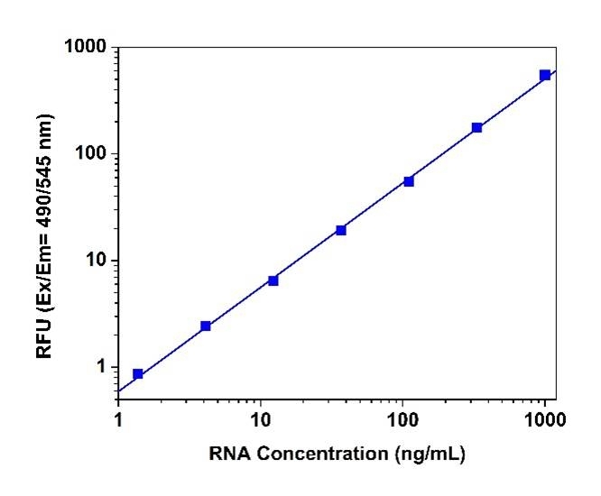
Detecting and quantitating small amounts of RNA is extremely important for a wide variety of molecular biology procedures such as measuring yields of in vitro transcribed RNA and measuring RNA concentrations before performing Northern blot analysis, S1 nuclease assays, RNase protection assays, cDNA library preparation, reverse transcription PCR, and differential display PCR. The most commonly used technique for measuring nucleic acid concentration is the determination of absorbance at 260 nm. The major disadvantage of the absorbance-based method is the interferences caused by proteins, free nucleotides and other UV absorbing compounds. The use of sensitive, fluorescent nucleic acid stains alleviates this interference problem. StrandBrite™ RNA quantifying reagent is an ultrasensitive fluorescent nucleic acid stain for quantitating RNA in solution. StrandBrite™ RNA quantifying reagent can detect as little as 5 ng/mL RNA with a fluorescence microplate reader or fluorometer. Our StrandBrite™ Green Fluorimetric RNA Quantitation Kit includes our StrandBrite™ Green nucleic acid stain with an optimized and robust protocol. It provides a convenient method for quantifying RNA in solutions.

Platform


Spectrofluorometer

Excitation490 nm
Emission545 nm
Cutoff515 nm

Components


Example protocol


AT A GLANCE

Protocol Summary
  1. Add 1 mL RNA standards or test samples in each cuvette
  2. Add 1 mL StrandBrite Green™ working solution
  3. Incubate at RT for 2-5 minutes
  4. Monitor the fluorescence at Ex/Em=490/545 nm 
Important      The following protocol is an example for quantifying RNA with StrandBrite™ Green. Allow all the components to warm to room temperature before opening. Prevent RNase contamination of the StrandBrite™ reagent and kit components. Always use clean disposable gloves while handling all materials. Use nuclease-free water, and sterile, disposable polypropylene plastic ware for reagent preparation. No data are available for addressing the mutagenicity or toxicity of StrandBrite™ Green RNA stain. Because this reagent binds to nucleic acids, it should be treated as a potential mutagen and handled with appropriate care. The DMSO stock solution should be handled with particular caution as DMSO is known to facilitate the entry of organic molecules into tissues.

PREPARATION OF STOCK SOLUTIONS

Unless otherwise noted, all unused stock solutions should be divided into single-use aliquots and stored at -20 °C after preparation. Avoid repeated freeze-thaw cycles.

Assay Buffer stock solution (1X)
Prepare a 1X Assay Buffer stock solution by diluting the concentrated 10X Assay Buffer (Component B) with sterile, distilled, nuclease-free water.

PREPARATION OF STANDARD SOLUTION

For convenience, use the Serial Dilution Planner:
https://www.aatbio.com/tools/serial-dilution/17656


RNA standard
Add 20 µL of 100 µg/mL RNA stock solution (Component C) to 1.98 mL of 1X Assay Buffer to have 1 µg/mL RNA solution, and then perform 1:3 serial dilutions to get remaining serially diluted RNA standards. Note: Unused Ribosomal RNA Standard (Component C) should be divided into single use aliquots in nuclease-free plastic vials and stored at -20°C.

PREPARATION OF WORKING SOLUTION

Prepare StrandBrite™ Green working solution by making a 200-fold dilution of the concentrated DMSO solution in 1X assay buffer. For example, to prepare enough working solution to assay 10 samples in a 2 mL final volume, add 50 μL of StrandBrite™ Green (Component A) into 10 mL of 1X assay buffer. Protect the working solution from light by covering it with foil or placing it in the dark.
Note     We recommend preparing this solution in a plastic container rather than glass, as the dye may adsorb to glass surfaces. For best results, this solution should be used within a few hours of its preparation.

SAMPLE EXPERIMENTAL PROTOCOL

  1. Add 1 mL of StrandBrite™ Green working solution to each cuvette containing 1 mL of the RNA standard, blank control, and test samples to make the total RNA assay volume of 2 mL/cuvette.
  2. Incubate the reaction at room temperature for 2 to 5 minutes, protected from light.
  3. Monitor the fluorescence increase with a spectrofluorometer at Ex/Em = 490/545 nm.
    Note     To minimize photobleaching effects, keep the time for fluorescence measurement constant for all samples. 

Spectrum


Open in Advanced Spectrum Viewer
spectrum

Spectral properties

Excitation (nm)509
Emission (nm)527

Images


Citations


View all 2 citations: Citation Explorer
The nucleolar shell provides anchoring sites for DNA untwisting
Authors: Fukute, Jumpei and Maki, Koichiro and Adachi, Taiji
Journal: Communications Biology (2024): 83
Anticancer Activity of Reconstituted Ribonuclease S-Decorated Artificial Viral Capsid
Authors: Liang, Yingbing and Furukawa, Hiroto and Sakamoto, Kentarou and Inaba, Hiroshi and Matsuura, Kazunori
Journal: ChemBioChem (2022): e202200220

References


View all 31 references: Citation Explorer
Inhibitors of Streptococcus pneumoniae surface endonuclease EndA discovered by high-throughput screening using a PicoGreen fluorescence assay
Authors: Peterson EJ, Kireev D, Moon AF, Midon M, Janzen WP, Pingoud A, Pedersen LC, Singleton SF.
Journal: J Biomol Screen (2013): 247
Validation of a PicoGreen-based DNA quantification integrated in an RNA extraction method for two-dimensional and three-dimensional cell cultures
Authors: Chen Y, Sonnaert M, Roberts SJ, Luyten FP, Schrooten J.
Journal: Tissue Eng Part C Methods (2012): 444
Characterization of PicoGreen interaction with dsDNA and the origin of its fluorescence enhancement upon binding
Authors: Dragan AI, Casas-Finet JR, Bishop ES, Strouse RJ, Schenerman MA, Geddes CD.
Journal: Biophys J (2010): 3010
Comparison of SYBR Green I-, PicoGreen-, and [3H]-hypoxanthine-based assays for in vitro antimalarial screening of plants from Nigerian ethnomedicine
Authors: Abiodun OO, Gbotosho GO, Ajaiyeoba EO, Happi CT, Hofer S, Wittlin S, Sowunmi A, Brun R, Oduola AM.
Journal: Parasitol Res (2010): 933
Metal-enhanced PicoGreen fluorescence: application to fast and ultra-sensitive pg/ml DNA quantitation
Authors: Dragan AI, Bishop ES, Casas-Finet JR, Strouse RJ, Schenerman MA, Geddes CD.
Journal: J Immunol Methods (2010): 95
Quantification of dsDNA using the Hitachi F-7000 Fluorescence Spectrophotometer and PicoGreen dye
Authors: Moreno LA, Cox KL.
Journal: J Vis Exp. (2010)
Factors affecting quantification of total DNA by UV spectroscopy and PicoGreen fluorescence
Authors: Holden MJ, Haynes RJ, Rabb SA, Satija N, Yang K, Blasic JR, Jr.
Journal: J Agric Food Chem (2009): 7221
Development and characterization of a novel host cell DNA assay using ultra-sensitive fluorescent nucleic acid stain "PicoGreen"
Authors: Ikeda Y, Iwakiri S, Yoshimori T.
Journal: J Pharm Biomed Anal (2009): 997
Enhanced DNA dynamics due to cationic reagents, topological states of dsDNA and high mobility group box 1 as probed by PicoGreen
Authors: Noothi SK, Kombrabail M, Kundu TK, Krishnamoorthy G, Rao BJ.
Journal: FEBS J (2009): 541
Label-free DNA sequence detection with enhanced sensitivity and selectivity using cationic conjugated polymers and PicoGreen
Authors: Ren X, Xu QH.
Journal: Langmuir (2009): 43