logo
AAT Bioquest

ThiolTrace™ Violet 500

The comparison in the fluorescence intensity of ThiolTrace&trade; Violet 500 with ThiolTracker<sup>TM</sup> Violet (Thermo Scientific) in Jurkat cells in the <strong>presence of cell culture medium</strong>. Jurkat cells were dye loaded with ThiolTrace&trade; Violet 500 or ThiolTracker<sup>TM</sup> Violet for 20 minutes in a 37 <sup>o</sup>C, 5% CO<sub>2</sub> incubator. The fluorescence intensity was measured using ACEA NovoCyte 3000 flow cytometer with Pacific Orange channel.
Ordering information
Price
Catalog Number
Unit Size
Quantity
Add to cart
Additional ordering information
Telephone1-800-990-8053
Fax1-800-609-2943
Emailsales@aatbio.com
InternationalSee distributors
Bulk requestInquire
Custom sizeInquire
ShippingStandard overnight for United States, inquire for international
Request quotation
Physical properties
Molecular weight557.01
SolventDMSO
Spectral properties
Excitation (nm)415
Emission (nm)499
Storage, safety and handling
H-phraseH303, H313, H333
Hazard symbolXN
Intended useResearch Use Only (RUO)
R-phraseR20, R21, R22
StorageFreeze (< -15 °C); Minimize light exposure
UNSPSC12352200

OverviewpdfSDSpdfProtocol


Molecular weight
557.01
Excitation (nm)
415
Emission (nm)
499
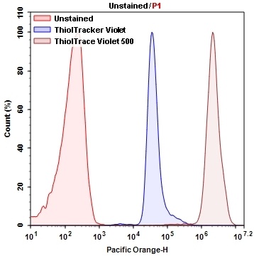
The subcellular detection and localization of GSH is important in understanding the modulation of redox status, the effect of drugs, and the mechanisms of detoxification. ThiolTrace™ Violet 500 is a brighter and more robust intracellular thiol probe for monitoring intracelluar GSH than the commonly used mBBr, mBCl or Thiotracker™ Violet. Since reduced glutathione represents the majority of intracellular free thiols in the cell, ThiolTrace™ Violet 500 can be used in estimating the cellular level of reduced glutathione. It is at least 10X brighter than mBCL and other intracellular common thiol detection probes (e.g., Thiotracker Violet), and can be excited with UV or 405 nm excitation with a large Stokes shift. It can be fixed with aldehydes and permeabilized by Triton® X-100 (0.5%). It may be used in multiplex assays including cytotoxicity studies. ThiolTrace Violet 500 provides a simple, sensitive and reproducible tool to detect reduced GSH content in biological samples. ThiolTrace Violet 500 reacts with thiol to emit a strong fluorescence of 520-530 nm with excitation at the common violet 405 nm laser. When compared with ThiolTracker Violet (Thermo Fisher Scientific), ThiolTrace Violet 500 has 10-100 fold higher intensity in cell culture medium containing growth factors. ThiolTrace Violet 500 is compatible with wide variety of diluents including serum containing cell culture medium. ThiolTrace™ Violet 500 can be used for Flow Cytometry, HCS imaging and epifluorescent microscopy.

Platform


Flow cytometer

Excitation405 nm laser
Emission525/50 nm filter
Instrument specification(s)Pacific Orange channel

Fluorescence microscope

Excitation405 nm
Emission525 nm
Recommended plateBlack wall/clear bottom
Instrument specification(s)TRITC filterset

Example protocol


AT A GLANCE

Protocol Summary
  1. Prepare cells with test compounds at a density of 5 × 10⁵ to 1 × 10⁶ cells/mL
  2. Prepare and add ThiolTraceTM Violet 500 working solution to cells
  3. Incubate at 37oC for 20 to 30 minutes
  4. Read fluorescence intensity at Ex/Em = 405/525 nm-Pacific Orange filter set
Important Note

Thaw at room temperature before starting the experiment.
Note         For flow cytometry and fluorescence microscopy, 200 and 500 tests can be performed with the quantity provided, respectively.

CELL PREPARATION

For guidelines on cell sample preparation, please visit https://www.aatbio.com/resources/guides/cell-sample-preparation.html

PREPARATION OF STOCK SOLUTIONS

Unless otherwise noted, all unused stock solutions should be divided into single-use aliquots and stored at -20 °C after preparation. Avoid repeated freeze-thaw cycles

ThiolTraceTM Violet 500 stock solution (500X)

Add 200 µL of DMSO (Not provided) into the vial of ThiolTraceTM Violet 500, and mix well.
Note         Aliquot and stored the unused ThiolTraceTM Violet 500 stock solution at -20 oC. Avoid repeated freeze/thaw cycles.

PREPARATION OF WORKING SOLUTION

ThiolTraceTM Violet 500 working solution (1X)

Add 1 µL of ThiolTraceTM Violet 500 stock solution into 0.5 mL of buffer of your choice, and mix well.
Note         ThiolTraceTM Violet 500 working solution can be prepared in the cell culture medium without serum.

SAMPLE EXPERIMENTAL PROTOCOL

  1. Treat cells with test compounds for a desired period of time.

    Note         For adherent cells, gently lift the cells with 0.5 mM EDTA to keep the cells intact, and wash the cells once with serum-containing media prior to the incubation with ThiolTrace™ Violet working solution.

    Note         The appropriate incubation time depends on the individual cell type and cell concentration used. Optimize the incubation time for each experiment.
  2. Centrifuge the cells at 1000 rpm for 4 minutes, and wash cells in 1 mL of buffer of your choice (Optional).
  3. Resuspend cells in 0.5 mL ThiolTraceTM Violet 500 working solution and incubate them at 37oC incubator for 20 to 30 minutes.

    Note         For the fluorescence microscopy, add 200 µL of the ThiolTraceTM Violet 500 working solution per well.
  4. Centrifuge the cells at 1000 rpm for 4 minutes, and then wash cells in 1 mL of buffer of your choice (Optional).
  5. Resuspend in buffer and monitor the fluorescence intensity with a flow cytometer using Pacific Orange filter set (Ex/Em = 405/525 nm). Gate on the cells of interest, excluding debris.

Calculators


Common stock solution preparation

Table 1. Volume of DMSO needed to reconstitute specific mass of ThiolTrace™ Violet 500 to given concentration. Note that volume is only for preparing stock solution. Refer to sample experimental protocol for appropriate experimental/physiological buffers.

0.1 mg0.5 mg1 mg5 mg10 mg
1 mM179.53 µL897.65 µL1.795 mL8.976 mL17.953 mL
5 mM35.906 µL179.53 µL359.06 µL1.795 mL3.591 mL
10 mM17.953 µL89.765 µL179.53 µL897.65 µL1.795 mL

Molarity calculator

Enter any two values (mass, volume, concentration) to calculate the third.

Mass (Calculate)Molecular weightVolume (Calculate)Concentration (Calculate)Moles
/=x=

Spectrum


Open in Advanced Spectrum Viewer
spectrum

Spectral properties

Excitation (nm)415
Emission (nm)499

Product Family


Images


Citations


View all 12 citations: Citation Explorer
A highly sensitive two-photon fluorescent probe for glutathione with near-infrared emission at 719nm and intracellular glutathione imaging
Authors: Huang, C., Qian, Y.
Journal: Spectrochim Acta A Mol Biomol Spectrosc (2019): 68-76
Glutathione-driven Cu(i)-O2 chemistry: a new light-up fluorescent assay for intracellular glutathione
Authors: Gao, P. F., Mao, Y. T., Yang, T., Zou, H. Y., Li, Y. F., Huang, C. Z.
Journal: Analyst (2018): 2486-2490
Nitrogen-doped carbon quantum dots as a fluorescent probe to detect copper ions, glutathione, and intracellular pH
Authors: Liao, S., Huang, X., Yang, H., Chen, X.
Journal: Anal Bioanal Chem (2018): 7701-7710
Graphene Quantum Dot-MnO2 Nanosheet Based Optical Sensing Platform: A Sensitive Fluorescence &quot;Turn Off-On&quot; Nanosensor for Glutathione Detection and Intracellular Imaging
Authors: Yan, X., Song, Y., Zhu, C., Song, J., Du, D., Su, X., Lin, Y.
Journal: ACS Appl Mater Interfaces (2016): 21990-6
A sensitive turn-on fluorescent probe for intracellular imaging of glutathione using single-layer MnO2 nanosheet-quenched fluorescent carbon quantum dots
Authors: He, D., Yang, X., He, X., Wang, K., Yang, X., He, X., Zou, Z.
Journal: Chem Commun (Camb) (2015): 14764-7
A mitochondria-targetable fluorescent probe for dual-channel NO imaging assisted by intracellular cysteine and glutathione
Authors: Sun, Y. Q., Liu, J., Zhang, H., Huo, Y., Lv, X., Shi, Y., Guo, W.
Journal: J Am Chem Soc (2014): 12520-3
A fluorescent probe for intracellular cysteine overcoming the interference by glutathione
Authors: Tian, M., Guo, F., Sun, Y., Zhang, W., Miao, F., Liu, Y., Song, G., Ho, C. L., Yu, X., Sun, J. Z., Wong, W. Y.
Journal: Org Biomol Chem (2014): 6128-33
Designing an intracellular fluorescent probe for glutathione: two modulation sites for selective signal transduction
Authors: Isik, M., Guliyev, R., Kolemen, S., Altay, Y., Senturk, B., Tekinay, T., Akkaya, E. U.
Journal: Org Lett (2014): 3260-3
Turn-on fluorescence sensor for intracellular imaging of glutathione using g-C(3)N(4) nanosheet-MnO(2) sandwich nanocomposite
Authors: Zhang, X. L., Zheng, C., Guo, S. S., Li, J., Yang, H. H., Chen, G.
Journal: Anal Chem (2014): 3426-34
Detection of intracellular glutathione using ThiolTracker violet stain and fluorescence microscopy
Authors: M, undefined and avilli, B. S., Janes, M. S.
Journal: Curr Protoc Cytom (2010): Unit 9 35