Products
Services
Resources
Selection Guides
About
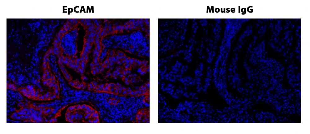
XFD532 tyramide
XFD532 Tyramide is a yellow fluorescent dye with an excitation/emission of 534/553 nm that is used for flow cytometry and microscopy applications.
- Tyramide Signal Amplification (TSA): Facilitates ultrasensitive detection of low-abundance targets, ideal for IHC and ICC applications
- Bright & Stable XFD532 Dye: Delivers high quantum yield with robust resistance to photobleaching and pH variations (4–10)
| Catalog | Size | Price | Quantity |
|---|---|---|---|
| 11072 | 200 Slides | Price |

Physical properties
| Molecular weight | 847.06 |
| Solvent | DMSO |
Spectral properties
| Correction factor (260 nm) | 0.24 |
| Correction factor (280 nm) | 0.09 |
| Extinction coefficient (cm -1 M -1) | 81000 |
| Excitation (nm) | 534 |
| Emission (nm) | 553 |
| Quantum yield | 0.61 1 |
Storage, safety and handling
| H-phrase | H303, H313, H333 |
| Hazard symbol | XN |
| Intended use | Research Use Only (RUO) |
| R-phrase | R20, R21, R22 |
| Storage | Freeze (< -15 °C); Minimize light exposure |
| UNSPSC | 12171501 |
Instrument settings
| Fluorescence microscope | |
| Excitation | Cy3/TRITC filter set |
| Emission | Cy3/TRITC filter set |
| Recommended plate | Black wall/clear bottom |
Contact us
| Telephone | |
| Fax | |
| sales@aatbio.com | |
| International | See distributors |
| Bulk request | Inquire |
| Custom size | Inquire |
| Technical Support | Contact us |
| Request quotation | Request |
| Purchase order | Send to sales@aatbio.com |
| Shipping | Standard overnight for United States, inquire for international |
Page updated on July 12, 2026