logo
AAT Bioquest

Xite™ Green beta-D-galactopyranoside

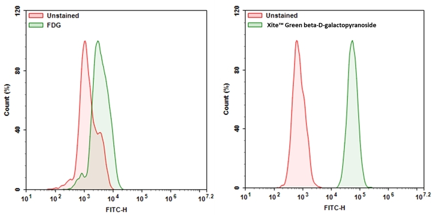
Expression of β-gal was measured with Xite™ Green beta-D-galactopyranoside. 9L-LacZ cells (cells that overexpressed β-gal) were incubated with Xite™ Green beta-D-galactopyranoside or FDG for 30 mins at 37 °C. The signal was acquired with FITC channel using a NovoCyte Flow Cytometer (ACEA Biosciences).
Ordering information
Price
Catalog Number
Unit Size
Quantity
Add to cart
Additional ordering information
Telephone1-800-990-8053
Fax1-800-609-2943
Emailsales@aatbio.com
InternationalSee distributors
Bulk requestInquire
Custom sizeInquire
ShippingStandard overnight for United States, inquire for international
Request quotation
Physical properties
Molecular weight494.50
SolventDMSO
Storage, safety and handling
H-phraseH303, H313, H333
Hazard symbolXN
Intended useResearch Use Only (RUO)
R-phraseR20, R21, R22
StorageFreeze (< -15 °C); Minimize light exposure
UNSPSC12171501

OverviewpdfSDSpdfProtocol


Molecular weight
494.50
Xite™ Green beta-D-galactopyranoside is a fluorogenic substrate for beta-galactosidase (β-gal). Compared to the existing beta-galactosidase substrates (e.g., the commonly used FDG), it has much better cell permeability. Xite™ Green beta-D-galactopyranoside readily enters cells where it gets cleaved by β-gal, producing Xite™ Green, a strongly fluorescent product. The strongly fluorescent Xite™ Green is well retained in cells, making it easy to be detected with a flow cytometer and fluorescence microscope. Xite™ Green beta-D-galactopyranoside provides a simple and sensitive tool to detect beta-galactosidase activity. Xite™ Green beta-D-galactopyranoside might be used as a simple tool for measuring cellular senescence in cells since β-gal has been identified as a reliable marker for cellular senescence.
Platform


Flow cytometer

Excitation488 nm laser
Emission530/30 nm filter
Instrument specification(s)FITC channel

Fluorescence microscope

ExcitationFITC filter set
EmissionFITC filter set
Recommended plateBlack wall/clear bottom

Example protocol


AT A GLANCE

Protocol summary
  1. Treat samples as desired
  2. Prepare and add Xite™ Green beta-D-galactopyranoside working solution to samples
  3. Incubate samples at 37 °C for 15 to 45 minutes
  4. Monitor the fluorescence intensity using flow cytometer with 530/30 nm filter (FITC channel) or using fluorescence microscopy with FITC filter set 

PREPARATION OF STOCK SOLUTIONS

Unless otherwise noted, all unused stock solutions should be divided into single-use aliquots and stored at -20 °C after preparation. Avoid repeated freeze-thaw cycles.

Xite™ Green beta-D-galactopyranoside stock solution
Add appropriate amount of DMSO into Xite™ Green beta-D-galactopyranoside to make 2-5 mM Xite™ Green beta-D-galactopyranoside stock solution. Note: Store the unused Xite™ Green beta-D-galactopyranoside stock solution at -20 °C in single use aliquots.

PREPARATION OF WORKING SOLUTION

Xite™ Green beta-D-galactopyranoside working solution
Prepare 1-20 µM of Xite™ Green beta-D-galactopyranoside working solution in buffer of your choice. Note: Xite™ Green beta-D-galactopyranoside working solution should be used promptly. Note: The concentration of the Xite™ Green beta-D-galactopyranoside should be optimized for different cell types and conditions.

SAMPLE EXPERIMENTAL PROTOCOL

The following protocol can be used as a guideline and should be optimized according to the needs.
  1. Treat your samples as desired.
  2. Remove the treatment and wash the cells with buffer of your choice such as DPBS. Note: For selectively tracking β-Gal in live cells, cells can be treated with Bafilomycin A1 for blocking endogenous β-Gal. Optimum concentration of Bafilomycin A1 may vary on type of cells.
  3. Add Xite™ Green beta-D-galactopyranoside working solution for 15-45 minutes and incubate the samples at 37 °C incubator. Note: Optimal time for incubation needs to be determined experimentally.
  4. Remove the working solution and wash cells with buffer of your choice.
  5. Resuspend the cells in buffer of your choice and monitor the fluorescence intensity with flow cytometer using 530/30 nm filter (FITC channel) or fluorescence microscope with FITC filter set. 

Calculators


Common stock solution preparation

Table 1. Volume of DMSO needed to reconstitute specific mass of Xite™ Green beta-D-galactopyranoside to given concentration. Note that volume is only for preparing stock solution. Refer to sample experimental protocol for appropriate experimental/physiological buffers.

0.1 mg0.5 mg1 mg5 mg10 mg
1 mM202.224 µL1.011 mL2.022 mL10.111 mL20.222 mL
5 mM40.445 µL202.224 µL404.449 µL2.022 mL4.044 mL
10 mM20.222 µL101.112 µL202.224 µL1.011 mL2.022 mL

Molarity calculator

Enter any two values (mass, volume, concentration) to calculate the third.

Mass (Calculate)Molecular weightVolume (Calculate)Concentration (Calculate)Moles
/=x=

Images


References


View all 18 references: Citation Explorer
Novel fluorescent probe for rapid and ratiometric detection of β-galactosidase and live cell imaging.
Authors: Chen, Xiangzhu and Zhang, Xueyan and Ma, Xiaodong and Zhang, Yuanyuan and Gao, Gui and Liu, Jingjing and Hou, Shicong
Journal: Talanta (2019): 308-313
Targeting senescence improves angiogenic potential of adipose-derived mesenchymal stem cells in patients with preeclampsia.
Authors: Suvakov, Sonja and Cubro, Hajrunisa and White, Wendy M and Butler Tobah, Yvonne S and Weissgerber, Tracey L and Jordan, Kyra L and Zhu, Xiang Y and Woollard, John R and Chebib, Fouad T and Milic, Natasa M and Grande, Joseph P and Xu, Ming and Tchkonia, Tamara and Kirkland, James L and Lerman, Lilach O and Garovic, Vesna D
Journal: Biology of sex differences (2019): 49
SA-β-Galactosidase-Based Screening Assay for the Identification of Senotherapeutic Drugs.
Authors: Fuhrmann-Stroissnigg, Heike and Santiago, Fernando E and Grassi, Diego and Ling, YuanYuan and Niedernhofer, Laura J and Robbins, Paul D
Journal: Journal of visualized experiments : JoVE (2019)
Cellular and cytoskeletal alterations of scleral fibroblasts in response to glucocorticoid steroids.
Authors: Bogarin, Thania and Saraswathy, Sindhu and Akiyama, Goichi and Xie, Xiaobin and Weinreb, Robert N and Zheng, Jie and Huang, Alex S
Journal: Experimental eye research (2019): 107774
Tumor cell escape from therapy-induced senescence.
Authors: Saleh, Tareq and Tyutyunyk-Massey, Liliya and Murray, Graeme F and Alotaibi, Moureq R and Kawale, Ajinkya S and Elsayed, Zeinab and Henderson, Scott C and Yakovlev, Vasily and Elmore, Lynne W and Toor, Amir and Harada, Hisashi and Reed, Jason and Landry, Joseph W and Gewirtz, David A
Journal: Biochemical pharmacology (2019): 202-212
Fluorescent probes for selective protein labeling in lysosomes: a case of α-galactosidase A.
Authors: Bohl, Cornelius and Pomorski, Adam and Seemann, Susanne and Knospe, Anne-Marie and Zheng, Chaonan and Krężel, Artur and Rolfs, Arndt and Lukas, Jan
Journal: FASEB journal : official publication of the Federation of American Societies for Experimental Biology (2017): 5258-5267
Identification of a β-galactosidase transgene that provides a live-cell marker of transcriptional activity in growing oocytes and embryos.
Authors: Edwards, Nicole and Farookhi, Riaz and Clarke, Hugh J
Journal: Molecular human reproduction (2015): 583-93
Characterization of functional capacity of adult ventricular myocytes in long-term culture.
Authors: Liu, Shi J
Journal: International journal of cardiology (2013): 1923-36
Abrupt and dynamic changes in gene expression revealed by live cell arrays.
Authors: Walling, Maureen A and Shi, Hua and Shepard, Jason R E
Journal: Analytical chemistry (2012): 2737-44
Oxidative stress and cell senescence combine to cause maximal renal tubular epithelial cell dysfunction and loss in an in vitro model of kidney disease.
Authors: Small, David M and Bennett, Nigel C and Roy, Sandrine and Gabrielli, Brian G and Johnson, David W and Gobe, Glenda C
Journal: Nephron. Experimental nephrology (2012): 123-30