logo
AAT Bioquest

Z-IETD-ProRed™ 620

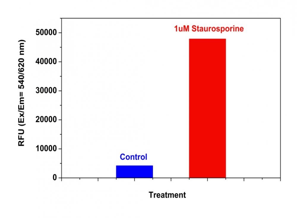
Detection of Caspase 8 Activity in Jurkat cells with Z-IETD-ProRed™ 620. Jurkat cells were seeded on the same day at 200,000 cells/90 µL/well in a Costar black wall/clear bottom 96-well plate. The cells were treated with staurosporine at the final concentration of 1 µM for 5 hours while the untreated cells were used as control. The caspase 8 assay solution (100 µL/well) was added and incubated at room temperature for 1 hour. The fluorescence intensity was measured at Ex/Em = 540/620 nm with a FlexStation™ microplate reader (Molecular Devices).
Ordering information
Price
Catalog Number
Unit Size
Quantity
Add to cart
Additional ordering information
Telephone1-800-990-8053
Fax1-800-609-2943
Emailsales@aatbio.com
InternationalSee distributors
Bulk requestInquire
Custom sizeInquire
ShippingStandard overnight for United States, inquire for international
Request quotation
Physical properties
Molecular weight1565.59
SolventDMSO
Spectral properties
Excitation (nm)532
Emission (nm)619
Storage, safety and handling
H-phraseH303, H313, H333
Hazard symbolXN
Intended useResearch Use Only (RUO)
R-phraseR20, R21, R22
StorageFreeze (< -15 °C); Minimize light exposure
UNSPSC12352200

OverviewpdfSDSpdfProtocol


See also: Caspases
Molecular weight
1565.59
Excitation (nm)
532
Emission (nm)
619
ProRed™-derived protease substrates are colorless and non-fluorescent. Cleavage of blocking protease-cleavable peptide residue by caspases generates the strongly red fluorescent ProRed™ that can be monitored fluorimetrically at ~620 nm with excitation of ~530 nm. ProRed™-derived caspase substrates are the most sensitive red indicators for the fluorimetric detection of various caspase activities. This IETD-ProRed™ substrate is specific for detecting caspase 8.
Example protocol


AT A GLANCE

Important notes

It is important to store at <-15 °C and should be stored in cool, dark place.

It can be used within 12 months from the date of receipt. 

SAMPLE EXPERIMENTAL PROTOCOL

Following protocol only provides a guideline, and should be modified according to your specific needs.

General Solution Caspase Assays Using AMC, AFC, pNA, R110 and ProRed Substrates

  1. Prepare a 10 mM stock solution in DMSO.

  2. Prepare a 2X caspase substrate (50 µM) assay solution as the following: 50 µL substrate stock solution, 100 µL DTT (1M), 400 µL EDTA (100 mM), 10 mL Tris Buffer (20 mM), pH =7.4.

  3. Mix equal volume of the caspase standards or samples with 2X caspase substrate assay solution, and incubate the solutions at room temperature for at least 1 hour.

  4. Monitor the fluorescence using a fluorescence microplate reader, or absorbance using an absorbance microplate reader.

Cell Caspase Assays Using Cell-Permeable FMK Caspase Probes

  1. Prepare a 2-5 mM stock solution in DMSO.

  2. Treat cells as desired.

  3. Prepare a 2X permeable caspase substrate (20 µM) assay solution by diluting the DMSO stock solution (from Step 2.1) in Hanks with 20 mM Hepes buffer (HHBS).

  4. Mix equal volume of the treated cells with 2X caspase substrate assay solution (from Step 2.3), and incubate the cells in a 37°C, 5% CO2 incubator for at least1 hour.

  5. Wash the cells with HHBS for at least once.

  6. Monitor the fluorescence intensity by a flow cytometer, a fluorescence microscope or a fluorescence microplate reader.

Cell Caspase Assays Using Cell-Permeable FMK Caspase Probes (For #13470-13476 only)

  1. Prepare a 250X stock solution by adding 50 µL DMSO into the vial.

  2. Treat cells as desired.

  3. Add 250 X DMSO stock solution into the cell solution at a 1:250 ratio (such as 2 µL to 500 µL cells), and incubate the cells in a 37°C, 5% CO2 incubator for 1 hour.

  4. Wash the cells with HHBS for at least once.

  5. Monitor the fluorescence intensity by flow cytometer, fluorescence microscopy or fluorescent microplate reader.

Calculators


Common stock solution preparation

Table 1. Volume of DMSO needed to reconstitute specific mass of Z-IETD-ProRed™ 620 to given concentration. Note that volume is only for preparing stock solution. Refer to sample experimental protocol for appropriate experimental/physiological buffers.

0.1 mg0.5 mg1 mg5 mg10 mg
1 mM63.874 µL319.368 µL638.737 µL3.194 mL6.387 mL
5 mM12.775 µL63.874 µL127.747 µL638.737 µL1.277 mL
10 mM6.387 µL31.937 µL63.874 µL319.368 µL638.737 µL

Molarity calculator

Enter any two values (mass, volume, concentration) to calculate the third.

Mass (Calculate)Molecular weightVolume (Calculate)Concentration (Calculate)Moles
/=x=

Spectrum


Open in Advanced Spectrum Viewer
spectrum

Spectral properties

Excitation (nm)532
Emission (nm)619

Product Family


NameExcitation (nm)Emission (nm)
Z-DEVD-ProRed™ 620532619
Z-LEHD-ProRed™ 620532619

Images


Citations


View all 1 citations: Citation Explorer
Degenerin channel activation causes caspase-mediated protein degradation and mitochondrial dysfunction in adult C. elegans muscle
Authors: Gaffney, Christopher J and Shephard, Freya and Chu, Jeff and Baillie, David L and Rose, Ann and Constantin-Teodosiu, Dumitru and Greenhaff, Paul L and Szewczyk, Nathaniel J
Journal: Journal of Cachexia, Sarcopenia and Muscle (2015)

References


View all 101 references: Citation Explorer
3,5-dibenzyloxy-4'-hydroxystilbene induces early caspase-9 activation during apoptosis in human K562 chronic myelogenous leukemia cells
Authors: Roslie H, Chan KM, Rajab NF, Velu SS, Kadir SA, Bunyamin I, Weber JF, Thomas NF, Majeed AB, Myatt G, Inayat-Hussain SH.
Journal: J Toxicol Sci (2012): 13
Dopaminochrome induces caspase-independent apoptosis in the mesencephalic cell line, MN9D
Authors: Linsenbardt AJ, Breckenridge JM, Wilken GH, Macarthur H.
Journal: J Neurochem. (2012)
Acacetin induces apoptosis in human T cell leukemia Jurkat cells via activation of a caspase cascade
Authors: Watanabe K, Kanno S, Tomizawa A, Yomogida S, Ishikawa M.
Journal: Oncol Rep (2012): 204
5-Phenylselenyl- and 5-methylselenyl-methyl-2'-deoxyuridine induce oxidative stress, DNA damage, and caspase-2-dependent apoptosis in cancer cells
Authors: Kim BM, Rode AB, Han EJ, Hong IS, Hong SH.
Journal: Apoptosis (2012): 200
High cytosolic free calcium level signals apoptosis through mitochondria-caspase mediated pathway in rat eggs cultured in vitro
Authors: Tripathi A, Chaube SK.
Journal: Apoptosis (2012): 439
The effect of low-level laser to apoptosis of chondrocyte and caspases expression, including caspase-8 and caspase-3 in rabbit surgery-induced model of knee osteoarthritis
Authors: Lin HD, He CQ, Luo QL, Zhang JL, Zeng DX.
Journal: Rheumatol Int (2012): 759
A caspase 8-based suicide switch induces apoptosis in nanobody-directed chimeric receptor expressing T cells
Authors: Khaleghi S, Rahbarizadeh F, Ahmadv and D, Rasaee MJ, Pognonec P.
Journal: Int J Hematol (2012): 434
Taurine protects rat testes against doxorubicin-induced oxidative stress as well as p53, Fas and caspase 12-mediated apoptosis
Authors: Das J, Ghosh J, Manna P, Sil PC.
Journal: Amino Acids (2012): 1839
Low-Dose Spironolactone Prevents Apoptosis Repressor With Caspase Recruitment Domain Degradation During Myocardial Infarction
Authors: Loan Le TY, Mardini M, Howell VM, Funder JW, Ashton AW, Mihailidou AS.
Journal: Hypertension. (2012)
Steroid Receptor Coactivator-interacting Protein (SIP) Inhibits Caspase-independent Apoptosis by Preventing Apoptosis-inducing Factor (AIF) from Being Released from Mitochondria
Authors: Wang D, Liang J, Zhang Y, Gui B, Wang F, Yi X, Sun L, Yao Z, Shang Y.
Journal: J Biol Chem (2012): 12612