LDS 651
Ordering information
| Price | |
| Catalog Number | |
| Unit Size | |
| Quantity |
Additional ordering information
| Telephone | 1-800-990-8053 |
| Fax | 1-800-609-2943 |
| sales@aatbio.com | |
| International | See distributors |
| Bulk request | Inquire |
| Custom size | Inquire |
| Shipping | Standard overnight for United States, inquire for international |
Physical properties
| Molecular weight | 473.40 |
| Solvent | DMSO |
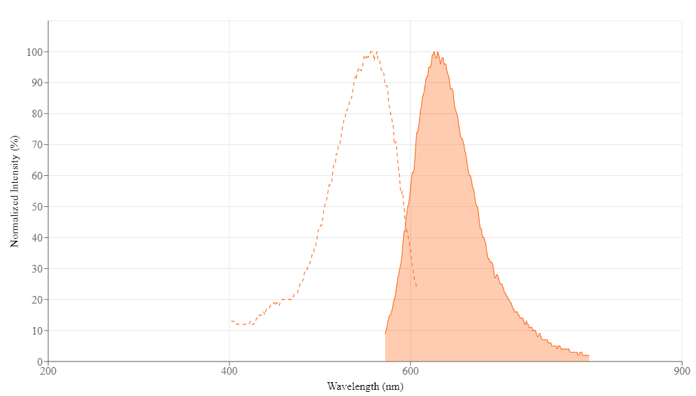
Spectral properties
| Excitation (nm) | 557 |
| Emission (nm) | 630 |
Storage, safety and handling
| H-phrase | H303, H313, H333 |
| Hazard symbol | XN |
| Intended use | Research Use Only (RUO) |
| R-phrase | R20, R21, R22 |
| Storage | Freeze (< -15 °C); Minimize light exposure |
| UNSPSC | 12171501 |
| Overview |
See also: Nucleus
Molecular weight 473.40 | Excitation (nm) 557 | Emission (nm) 630 |
LDS 651 is an analog of LDS 751. LDS 651 has almost identical intracellular staining pattern to LDS 751. LDS 651 is optimized to be excited by Violet Laser while LDS 751 is excited by Blue Laser at 488 nm. Both LDS 651 and LDS 751 are cell-permeant fluorescent dyes. Although both of LDS 651 and LDS 751 bind to DNA, they may be excluded from the nucleus and bind to other cellular structures such as the polarized membranes of mitochondria. Cautions need be taken when LDS 651 and LDS 751 are used for analyzing live cells and their fluorescence as being indicative of nuclear status. LDS 651 and LDS 751 might be used to separate red blood cells from nucleated cells.
Platform
Flow cytometer
| Excitation | 488 nm laser |
| Emission | 695/40 nm filter |
| Instrument specification(s) | PerCP channel |
Example protocol
PREPARATION OF STOCK SOLUTIONS
Unless otherwise noted, all unused stock solutions should be divided into single-use aliquots and stored at -20 °C after preparation. Avoid repeated freeze-thaw cycles.
Note Store the unused stock solution in small aliquots at -20 °C, protected from light.
LDS 651 stock solution
Prepare a 1 mM stock solution by adding the appropriate amount of DMSO.Note Store the unused stock solution in small aliquots at -20 °C, protected from light.
PREPARATION OF WORKING SOLUTION
LDS 651 working solution
Dilute the 1 mM LDS 651 stock solution to 1 to 10 µM LDS 651 working solution in the buffer of your choice.Note Prepare the working solution immediately before use.
Note The LDS 651 working solution should not be stored or reused.
Note The concentration of the LDS 651 working solution should be optimized for different cell types and conditions.
SAMPLE EXPERIMENTAL PROTOCOL
The following sample protocol is provided as a basic guide for the development of your own staining protocol. The concentrations of the reagents required for the optimal staining may vary depending on the density of cells (i.e. Leukocytes) and other materials in the sample.
- Add the LDS 651 working solution in samples.
- Incubate the samples at 37 °C for 5-10 minutes.
- Set a fluorescence threshold to detect cells stained positive with LDS-651, thus excluding erythrocytes and unbound single platelets from the display.
Calculators
Common stock solution preparation
Table 1. Volume of DMSO needed to reconstitute specific mass of LDS 651 to given concentration. Note that volume is only for preparing stock solution. Refer to sample experimental protocol for appropriate experimental/physiological buffers.
| 0.1 mg | 0.5 mg | 1 mg | 5 mg | 10 mg | |
| 1 mM | 211.238 µL | 1.056 mL | 2.112 mL | 10.562 mL | 21.124 mL |
| 5 mM | 42.248 µL | 211.238 µL | 422.476 µL | 2.112 mL | 4.225 mL |
| 10 mM | 21.124 µL | 105.619 µL | 211.238 µL | 1.056 mL | 2.112 mL |
Molarity calculator
Enter any two values (mass, volume, concentration) to calculate the third.
| Mass (Calculate) | Molecular weight | Volume (Calculate) | Concentration (Calculate) | Moles | ||||
| / | = | x | = |
Images

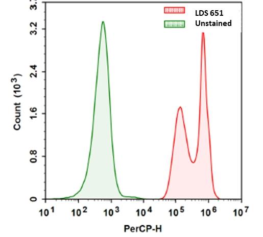
Figure 1. Flow cytometric analysis with LDS 651. Jurkat cells were stained with LDS 651 as per the protocol and response was measured with NovoCyte flow cytometer using PerCP channel.
References
View all 46 references: Citation Explorer
Quantifying permeabilization and activity recovery of Bacillus spores in adverse conditions for growth.
Authors: Trunet, C and Ngo, H and Coroller, L
Journal: Food microbiology (2019): 115-120
Authors: Trunet, C and Ngo, H and Coroller, L
Journal: Food microbiology (2019): 115-120
Calcium Ionophore, Calcimycin, Kills Leishmania Promastigotes by Activating Parasite Nitric Oxide Synthase.
Authors: Grekov, Igor and Pombinho, António R and Kobets, Tatyana and Bartůněk, Petr and Lipoldová, Marie
Journal: BioMed research international (2017): 1309485
Authors: Grekov, Igor and Pombinho, António R and Kobets, Tatyana and Bartůněk, Petr and Lipoldová, Marie
Journal: BioMed research international (2017): 1309485
Lipid bilayer properties control membrane partitioning, binding, and transport of p-glycoprotein substrates.
Authors: Clay, Adam T and Sharom, Frances J
Journal: Biochemistry (2013): 343-54
Authors: Clay, Adam T and Sharom, Frances J
Journal: Biochemistry (2013): 343-54
Interaction of LDS-751 with the drug-binding site of P-glycoprotein: a Trp fluorescence steady-state and lifetime study.
Authors: Lugo, Miguel R and Sharom, Frances J
Journal: Archives of biochemistry and biophysics (2009): 17-28
Authors: Lugo, Miguel R and Sharom, Frances J
Journal: Archives of biochemistry and biophysics (2009): 17-28
Flow cytometric assessment of homeostatic aging of reticulocytes in rats.
Authors: Wiczling, Paweł and Krzyzanski, Wojciech
Journal: Experimental hematology (2008): 119-27
Authors: Wiczling, Paweł and Krzyzanski, Wojciech
Journal: Experimental hematology (2008): 119-27
Determination of the formation of dark state via depleted spontaneous emission in a complex solvated molecule.
Authors: Guo, Xunmin and Wang, Sufan and Xia, Andong and Su, Hongmei
Journal: The journal of physical chemistry. A (2007): 5800-5
Authors: Guo, Xunmin and Wang, Sufan and Xia, Andong and Su, Hongmei
Journal: The journal of physical chemistry. A (2007): 5800-5
Comparison of sample fixation and the use of LDS-751 or anti-CD45 for leukocyte identification in mouse whole blood for flow cytometry.
Authors: Maes, Melissa L and Davidson, Lisa B and McDonagh, Paul F and Ritter, Leslie S
Journal: Journal of immunological methods (2007): 79-86
Authors: Maes, Melissa L and Davidson, Lisa B and McDonagh, Paul F and Ritter, Leslie S
Journal: Journal of immunological methods (2007): 79-86
A novel flow cytometric assay of human whole blood neutrophil and monocyte CD11b levels: upregulation by chemokines is related to receptor expression, comparison with neutrophil shape change, and effects of a chemokine receptor (CXCR2) antagonist.
Authors: Nicholson, Grant C and Tennant, Rachel C and Carpenter, Donald C and Sarau, Henry M and Kon, Onn Min and Barnes, Peter J and Salmon, Michael and Vessey, Rupert S and Tal-Singer, Ruth and Hansel, Trevor T
Journal: Pulmonary pharmacology & therapeutics (2007): 52-9
Authors: Nicholson, Grant C and Tennant, Rachel C and Carpenter, Donald C and Sarau, Henry M and Kon, Onn Min and Barnes, Peter J and Salmon, Michael and Vessey, Rupert S and Tal-Singer, Ruth and Hansel, Trevor T
Journal: Pulmonary pharmacology & therapeutics (2007): 52-9
Interaction of LDS-751 and rhodamine 123 with P-glycoprotein: evidence for simultaneous binding of both drugs.
Authors: Lugo, Miguel R and Sharom, Frances J
Journal: Biochemistry (2005): 14020-9
Authors: Lugo, Miguel R and Sharom, Frances J
Journal: Biochemistry (2005): 14020-9
Cell cycle synchronization of Cupriavidus necator by continuous phasing measured via flow cytometry.
Authors: Fritsch, M and Starruss, J and Loesche, A and Mueller, S and Bley, Th
Journal: Biotechnology and bioengineering (2005): 635-42
Authors: Fritsch, M and Starruss, J and Loesche, A and Mueller, S and Bley, Th
Journal: Biotechnology and bioengineering (2005): 635-42
Application notes
FAQ
Are Cell Navigator® Cell Plasma Membrane Staining Kits suitable for cell culture medium samples?
Are mRNAs found in prokaryotes differ from those of eukaryotes?
Are spliceosomes associated with any diseases?
Are there any alternatives for ethidium bromide in agarose gels?
Are there any alternatives to Cy5?
Are mRNAs found in prokaryotes differ from those of eukaryotes?
Are spliceosomes associated with any diseases?
Are there any alternatives for ethidium bromide in agarose gels?
Are there any alternatives to Cy5?