Nucleic Acid Detection, Quantification and Imaging
In this issue
Latest News and Events: 69th AACC Annual Scientific Meeting
Buccutite™ Conjugation Kits: Quick and Easy Antibody Labeling
NAADP-AM, A New Player in Calcium Signaling Pathways
Nucleic Acid Detection, Quantification and Imaging
Quantitative Analysis of Thiols and Maleimides
AssayWise Letters 2017, Vol. 6(1)
Nucleic acid staining is an important tool that gives insight into cellular mechanisms and functionality of macromolecules like DNA and RNA. This is critical as these nucleic acids and their associated organelles are responsible for the development, maintenance and growth of living organisms. The combination of nucleic acid staining with applications such as fluorescence microscopy and flow cytometry also allow for spatiotemporal imaging, providing a powerful means for visualizing stained nucleic components in fixed and live cells.
Analyzing nucleic acid stains in cells provides an excellent overview of the cellular localization and organization of nucleic macromolecules and organelles with respect to time. Fixed cell stains in tandem with flow cytometry provide a snapshot of the distribution of nucleic macromolecules in a population of cells with respect to a single temporal time point. For example, DNA staining of fixed cells can be used to analyze the relationship between membrane integrity and the degradation of DNA at specific times during cellular events such as apoptosis. Nucleic acid staining in live cells for fluorescence microscopy aids in visualizing and understanding the spatiotemporal dynamics and organization of nucleic acids and organelles in real time. The observation of dynamic changes provides more insight into the operations of nucleic acids as they move within their environment during specific cellular functions, such as the regulation of gene expression. However, differentiating between nucleic macromolecules is considerably challenging and may prove difficult in studies where selective staining of nucleic acids is imperative for spatiotemporal analysis.
Obstacles associated with selectively staining nucleic acids are the structural and chemical similarities shared between DNA and ribonucleic acid (RNA). Both categories of nucleic acids are nearly identical, and therefore many staining techniques are applicable and synonymous for both. This non-specificity for staining nucleic acids can be detrimental to the signal quality of interest because of the impeding background noise from other nucleic molecules. For example, DNA imaging and localization studies may exhibit impeding background interference by RNA because of the non-specific binding of dye-conjugated nucleic acid probes. To address this issue, research has led to the improvement and development of sensitive tools and alternative techniques optimized to specifically target and stain DNA. A simple improvement to DNA staining involves the removal of RNA by pretreating the nucleic acid sample with RNAse, an enzyme that catalyzes the degradation of RNA into smaller components. Some improvements utilize cell-permeable dyes designed to selectively target specific configurations of DNA, such as the minor groove region of double helix DNA. Other improvements combine applications, such as gel electrophoresis and nucleic acid staining, to separate and stain large double-stranded DNA (dsDNA) macromolecules of a biological sample.
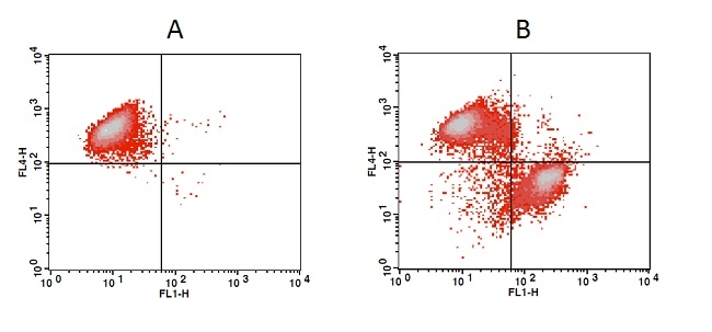
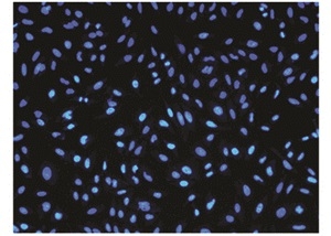
Image of dead cells stained with Nuclear Blue™ DCS1 (Cat# 17548). Fixed HeLa cells were plated on a 96-well microtiter plate, incubated with 2.5 nM Nuclear Blue™ DCS1 for 230 minutes and imaged with DAPI channel.
Obstacles associated with selectively staining nucleic acids are the structural and chemical similarities shared between DNA and ribonucleic acid (RNA). Both categories of nucleic acids are nearly identical, and therefore many staining techniques are applicable and synonymous for both. This non-specificity for staining nucleic acids can be detrimental to the signal quality of interest because of the impeding background noise from other nucleic molecules. For example, DNA imaging and localization studies may exhibit impeding background interference by RNA because of the non-specific binding of dye-conjugated nucleic acid probes. To address this issue, research has led to the improvement and development of sensitive tools and alternative techniques optimized to specifically target and stain DNA. A simple improvement to DNA staining involves the removal of RNA by pretreating the nucleic acid sample with RNAse, an enzyme that catalyzes the degradation of RNA into smaller components. Some improvements utilize cell-permeable dyes designed to selectively target specific configurations of DNA, such as the minor groove region of double helix DNA. Other improvements combine applications, such as gel electrophoresis and nucleic acid staining, to separate and stain large double-stranded DNA (dsDNA) macromolecules of a biological sample.

The increase in fluorescence intensity of Nuclear Green™ DCS1 (Cat# 17550) with the addition of camptothecin in Jurkat cells. Jurkat cells were treated overnight without (A) or with 20 nM camptothecin (B) in a 37 °C, 5% CO2 incubator, and then dye loaded with Nuclear Green™ DCS1 for 60 minutes. At the end of 15 minutes of Nuclear Green™ DCS1 dye loading, MitoLite™ NIR was added for multicolor analysis. The fluorescence intensity of Nuclear Green™ DCS1 and MitoLite™ NIR was measured with a FACSCalibur (Becton Dickinson) flow cytometer using FL1 channel (Nuclear Green™ DCS1) and FL4 channel (MitoLite™ NIR).
DNA Imaging

Image of a dividing (anaphase stage) human cancer cell taken with an epifluorescence microscope. The cancer cell was fixed and the DNA was stained with DAPI (blue), INCENP stained with GFP (green) and Tubulin stained red.
DNA imaging of cells can provide insight into the spatiotemporal dynamics of DNA during biological functions. For example, imaging can be used to visualize how the components of DNA move in relationship to one another during cell division. The common classes of commercially available nucleic acids stains are intercalating dyes and minor-groove binders.
Intercalating Dyes and Gel Electrophoresis

DNA molecular weight ladders stained with Cyber Green™ (Cat# 17590) and SYBR® Green I Nucleic Acid Gel Stain.
Ethidium bromide (EtBr) is a commonly used intercalating agent in molecular biology as a fluorescent dye for tagging and visualizing nucleic acids in gel electrophoresis. Its flat structure allows EtBr to intercalate, or insert, between nitrogenous bases in DNA molecules. Upon exposure to UV light, EtBr fluoresces, providing a means to visualize DNA molecules. EtBr's intercalating properties makes it a considerably potent mutagen that can interfere with the functionality of DNA molecules. Because of its high toxicity and mutagenic properties, many labs shy away from using EtBr as a DNA stain. To address toxicity concerns, AAT Bioquest offers a safer set of DNA dyes, Cyber Green™ (Cat# 17590) and Cyber Orange™ (Cat# 17595), that are excellent for nucleic acid gel stains. These dyes are commercially available individually as well as conveniently packaged in AAT Bioquest's Gelite™ Green and Gelite™ Orange Nucleic Acid Gel Staining Kits (Cat# 17589 and Cat# 17594). Gelite™ kits include all the necessary components, proprietary dyes and loading buffers combined with an optimized and robust protocol to effectively visualize nucleic acid gel stains.

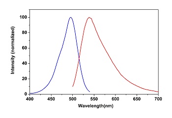
The excitation and emission spectra of Cyber Green™ Nucleic Acid Gel Stain (LEFT) and Cyber Orange™ Nucleic Acid Gel Stain (RIGHT) in the presence of calf thymus DNA.
Minor-Groove Binders and Fluorescence Microscopy
The structural significance of double stranded DNA provides another avenue for nucleic acid staining. Double helix DNA molecules have distinct chemical features on their molecular surface that act as recognition sites for small molecules and binding proteins. These sites are characterized by either shallow and wide major grooves, or deep and narrow minor grooves. Transcription factor proteins, essential for the regulation of gene expression, bind to the base pairs of the major grooves in dsDNA. Compounds that bind to the minor grooves in double helix DNA molecules are small fluorescent stains such as DAPI (Cat# 17507) and Hoechst stains (Cat# 17520, Cat# 17530 and Cat# 17537).


Left: Nuclei containing DNA was stained for fluorescence microscopy using Hoechst 33342 (Cat# 17530). Hoechst 33342 can be excited by UV light at 350 nm and will emit cyan fluorescence light with an emission max at 461 nm. Right: DAPI (Cat# 17507), a minor groove binding dye which binds to A-T rich regions of dsDNA, was used to stain and visualize cell nuclei containing DNA.
DAPI (4', 6-diamidino-2phenylindole) and Hoechst stains are cell permeable fluorescent dyes for labelling and visualizing DNA in fluorescence microscopy and flow cytometry. After permeating the cell membrane, these dyes have a strong affinity for A-T rich regions of the minor grooves in DNA. DAPI has an excitation of 358 nm and an emission of 461 nm, and Hoechst stains have a nearly identical excitation of 352 nm and emission of 461 nm. The blue emission of both dyes is convenient for multiplexing assays due to the minimal fluorescent overlap between DAPI or Hoechst stains with traditional green-fluorescent molecules.
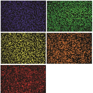
A composition of fluorescence images of HeLa cells stained with AAT Bioquest's Nuclear LCS1 dye series. HeLa cells were stained with Nuclear Violet™ (17543), Nuclear Green™ (Cat# 17540), Nuclear Red™ (Cat# 17542), Nuclear Yellow (Cat# 17539) and Nuclear Orange™ (Cat# 17541) LCS1 dyes in a Costar black wall/clear bottom 96-well microtiter plate.
Minor groove binders and intercalating dyes, discussed previously, are conventional approaches for imaging and analyzing localization of nucleic acids in cells. Utilization of these dyes in conjunction with imaging and localization studies has confirmed DNA organization and concentration to mitochondrial organelles and to the cell nucleus. For a more comprehensive understanding of the functionality of nucleic macromolecules and organelles, RNA staining is crucial. However, differentiating between RNA and DNA is difficult because of their similar composition. As a result, there has been an increase in interest regarding the research and development of RNA staining techniques. Modifications to dye-conjugated probes and DNA staining techniques can be altered to selectively target and stain cytoplasmic RNA molecules and organelles. Imaging and localization of RNA can provide vital information regarding its functionality in the cellular process of transcription and translation required for protein synthesis.
Additional Information
Table 1. Nucleic Acid Staining Reagents and Assay Kits
| Cat No. ▲ ▼ | Product Name ▲ ▼ | Ex (nm) ▲ ▼ | Em (nm) ▲ ▼ | Unit Size ▲ ▼ |
| 17590 | Helixyte™ Green Nucleic Acid Gel Stain *10,000X DMSO Solution* | 498 | 522 | 100 uL |
| 17590 | Helixyte™ Gold Nucleic Acid Gel Stain *10,000X DMSO Solution* | 496 | 539 | 1 mL |
| 17507 | DAPI [4,6-Diamidino-2-phenylindole, dihydrochloride] *10 mM solution in water* | 358 | 461 | 2 mL |
| 17589 | Gelite™ Green Nucleic Acid Gel Staining Kit | 497 | 520 | 1 Kit |
| 17594 | Gelite™ Orange Nucleic Acid Gel Staining Kit | 497 | 520 | 1 Kit |
| 17520 | Hoechst 33258 *CAS 23491-45-4* | 352 | 461 | 100 mg |
| 17530 | Hoechst 33342 *Ultrapure Grade* | 350 | 461 | 100 mg |
| 17537 | Hoechst 34580 *CAS 911004-45-0* | 368 | 437 | 5 mg |
| 17548 | Nuclear Blue™ DCS1 *5 mM DMSO Solution* | 350 | 461 | 0.5 mL |
| 17550 | Nuclear Green™ DCS1 *5 mM DMSO Solution* | 503 | 526 | 0.5 mL |
Further Reading
- Bao, Gang, Won Jong Rhee, and Andrew Tsourkas. "Fluorescent Probes for Live-Cell RNA Detection." Annual Review of Biomedical Engineering 11.1 (2009): 25-47.
- Brandenburg, Boerries, Lily Y. Lee, Melike Lakadamyali, Michael J. Rust, Xiaowei Zhuang, and James M. Hogle. "Imaging Poliovirus Entry in Live Cells." PLoS Biology 5.7 (2007): n. pag.
- Li, Qian, Yunkyung Kim, Joshua Namm, Amita Kulkarni, Gus R. Rosania, Young-Hoon Ahn, and Young-Tae Chang. "RNA-Selective, Live Cell Imaging Probes for Studying Nuclear Structure and Function." Chemistry & Biology 13.6 (2006): 615-23.
- Li, J. J. "Using molecular beacons as a sensitive fluorescence assay for enzymatic cleavage of single-stranded DNA." Nucleic Acids Research 28.11 (2000): n. pag.
- Liu, Zhe, Luke D. Lavis, and Eric Betzig. "Imaging Live-Cell Dynamics and Structure at the Single-Molecule Level." Molecular Cell 58.4 (2015): 644-59.
- Marras, Salvatore A.e., Sanjay Tyagi, and Fred Russell Kramer. "Real-time assays with molecular beacons and other fluorescent nucleic acid hybridization probes." Clinica Chimica Acta 363.1-2 (2006): 48-60.
- Mart-, Angel A., Steffen Jockusch, Nathan Stevens, Jingyue Ju, and Nicholas J. Turro. "Fluorescent Hybridization Probes for Sensitive and Selective DNA and RNA Detection." Accounts of Chemical Research 40.6 (2007): 402-09.
- Panning, Margaret M., and David M. Gilbert. "Spatio-temporal organization of DNA replication in murine embryonic stem, primary, and immortalized cells." Journal of Cellular Biochemistry 95.1 (2005): 74-82.
- Proudnikov, D., and A. Mirzabekov. "Chemical Methods of DNA and RNA Fluorescent Labeling." Nucleic Acids Research 24.22 (1996): 4535-542.
- Rye, Hays S., and Alexander N. Glazer. "Interaction of dimeric intercalating dyes with single-stranded DNA." Nucleic Acids Research 23.7 (1995): 1215-222.
- Santangelo, P. J. "Dual FRET molecular beacons for mRNA detection in living cells." Nucleic Acids Research 32.6 (2004): n. pag.
- Su, Xin, Xianjin Xiao, Chen Zhang, and Meiping Zhao. "Nucleic Acid Fluorescent Probes for Biological Sensing." Applied Spectroscopy 66.11 (2012): 1249-262.
- Urbanek, Martyna O., Paulina Galka-Marciniak, Marta Olejniczak, and Wlodzimierz J. Krzyzosiak. "RNA imaging in living cells — methods and applications." RNA Biology 11.8 (2014): 1083-095.
- Wark, Alastair W., Hye Jin Lee, and Robert M. Corn. "ChemInform Abstract: Multiplexed Detection Methods for Profiling MicroRNA Expression in Biological Samples." ChemInform 39.19 (2008): n. pag.