logo
AAT Bioquest

DAPI [4,6-Diamidino-2-phenylindole, dihydrochloride] *10 mM solution in water*

DAPI is a fluorescent stain that binds strongly to DNA. It is used extensively in fluorescence microscopy. Since DAPI passes through an intact cell membrane, it can be used to stain live cells besides fixed cells. For fluorescence microscopy, DAPI is excited with ultraviolet light. When bound to double-stranded DNA its absorption maximum is at 358 nm and its emission maximum is at 461 nm. One drawback of DAPI is that its emission is fairly broad. DAPI also binds to RNA although it is not as strongly fluorescent as it binds to DNA. Its emission shifts to around 500 nm when bound to RNA. DAPI's blue emission is convenient for multiplexing assays since there is very little fluorescence overlap between DAPI and green-fluorescent molecules like fluorescein and green fluorescent protein (GFP), or red-fluorescent stains like Texas Red. Besides labeling cell nuclei, DAPI is also used for the detection of mycoplasma or virus DNA in cell cultures.
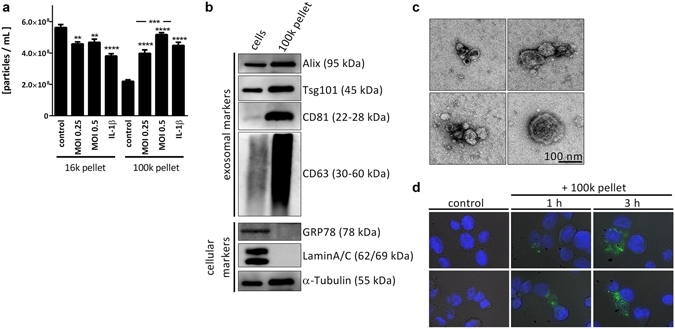
<em>L. pneumophila</em> infection increases the secretion of EVs in THP-1 cells. (a) Amount of EVs in response to <em>L. pneumophila</em> infection. THP-1 cells were treated with IL-1&beta; (1&thinsp;ng/mL) or infected with <em>L. pneumophila</em> (MOI 0.25 or 0.5, respectively) for 24&thinsp;h. NTA was performed with the distinct particle fractions separated by differential centrifugation. (b) Western blot for exosomal marker proteins. Whole cell lysate or 100&thinsp;k pellet derived from uninfected THP-1 cells were used. Equal protein amounts were loaded. (c) Transmission electron microscopy with 100&thinsp;k pellet. Purified EVs from THP-1 cells were fixed and visualized after negative staining with uranyl acetate. Scale bar represents 100&thinsp;nm. (d) Uptake of A549-derived EVs by THP-1 cells. A549 cells were stained with the membrane dye PKH67. EVs were collected (100&thinsp;k pellet) and incubated with THP-1 cells for 1 or 3&thinsp;h, respectively. Nuclei were stained by DAPI and pictures were taken with an original magnification of 630x. A: Data are shown as mean&thinsp;+&thinsp;SEM of three independent experiments. **p&thinsp;&lt;&thinsp;0.01, ***p&thinsp;&lt;&thinsp;0.001, ****p&thinsp;&lt;&thinsp;0.0001. B&ndash;D: representative results. Source: <strong><em>Legionella pneumophila</em> infection activates bystander cells differentially by bacterial and host cell vesicles </strong>by Jung et al., <em>Scientific Reports,</em> July 2017.
Gallery Image 2
Ordering information
Price
Unit size
Catalog Number17507
Quantity
Add to cart
Additional ordering information
Telephone1-800-990-8053
Fax1-800-609-2943
Emailsales@aatbio.com
InternationalSee distributors
Bulk requestInquire
Custom sizeInquire
ShippingStandard overnight for United States, inquire for international
Request quotation
Physical properties
Molecular weight350.25
SolventWater
Spectral properties
Extinction coefficient (cm -1 M -1)27000
Excitation (nm)359
Emission (nm)457
Storage, safety and handling
Certificate of OriginDownload PDF
H-phraseH303, H313, H340
Hazard symbolT
Intended useResearch Use Only (RUO)
R-phraseR20, R21, R68
StorageFreeze (< -15 °C); Minimize light exposure
UNSPSC41116134
CAS28718-90-3
Calculators

Common stock solution preparation

Table 1. Volume of Water needed to reconstitute specific mass of DAPI [4,6-Diamidino-2-phenylindole, dihydrochloride] *10 mM solution in water* to given concentration. Note that volume is only for preparing stock solution. Refer to sample experimental protocol for appropriate experimental/physiological buffers.

0.1 mg0.5 mg1 mg5 mg10 mg
1 mM285.51 µL1.428 mL2.855 mL14.276 mL28.551 mL
5 mM57.102 µL285.51 µL571.021 µL2.855 mL5.71 mL
10 mM28.551 µL142.755 µL285.51 µL1.428 mL2.855 mL

Molarity calculator

Enter any two values (mass, volume, concentration) to calculate the third.

Mass (Calculate)Molecular weightVolume (Calculate)Concentration (Calculate)Moles
/=x=
Spectrum
Citations
View all 50 citations: Citation Explorer
Deciphering the Role of Ferroptosis in Rheumatoid Arthritis: Synovial Transcriptome Analysis and Immune Infiltration Correlation
Authors: Wang, Hongli and Zhang, Miaomiao and Hu, Yiping and He, Juan and Zhong, Yuchao and Dai, Yong and Wang, Qingwen
Journal: Heliyon (2024)
The mesenchymal stromal cell (MSC) secretome promotes tissue regeneration and increases macrophage infiltration in acute and MRSA-infected skin wounds in vivo
Authors: Rajesh, Aarthi and Ju, Esther Da Eun and Oxford, Kelly A and Harman, Rebecca M and Van de Walle, Gerlinde R
Journal: Cytotherapy (2024)
Adipose Tissue and Translation Machinery in Metabolic Regulation
Authors: De Siqueira, Mirian Krystel
Journal: (2024)
Mechanosensitive membrane domains regulate calcium entry in arterial endothelial cells to protect against inflammation
Authors: Hong, Soon-Gook and Ashby, Julianne W and Kennelly, John P and Wu, Meigan and Steel, Michelle and Chattopadhyay, Eesha and Foreman, Rob and Tontonoz, Peter and Tarling, Elizabeth J and Turowski, Patric and others,
Journal: The Journal of Clinical Investigation (2024)
A novel HDAC8 inhibitor H7E exerts retinoprotective effects against glaucomatous injury via ameliorating aberrant M{\"u}ller glia activation and oxidative stress
Authors: Wu, Liang-Huan and Cheng, Yu-Wen and Lin, Fan-Li and Hsu, Kai-Cheng and Wang, Mong-Heng and Yen, Jing-Lun and Wang, Tsung-Jen and Lin, Tony Eight and Liu, Yi-Chien and Huang, Wei-Jan and others,
Journal: Biomedicine \& Pharmacotherapy (2024): 116538
References
View all 134 references: Citation Explorer
Reliable DNA ploidy determination in dehydrated tissues of vascular plants by DAPI flow cytometry--new prospects for plant research
Authors: Suda J, Travnicek P.
Journal: Cytometry A (2006): 273
Identification of all pachytene bivalents in the common shrew using DAPI-staining of synaptonemal complex spreads
Authors: Belonogova NM, Karamysheva TV, Biltueva LS, Perepelov EA, Minina JM, Polyakov AV, Zhdanova NS, Rubtsov NB, Searle JB, Borodin PM.
Journal: Chromosome Res (2006): 673
Physical localization of ribosomal genes and chromosome DAPI banding by in situ hybridization in Medicago sativa L
Authors: Chen JM, Hong YH, Wang YP, Bowley S, Wan JM.
Journal: Yi Chuan (2006): 184
DNA staining with the fluorochromes EtBr, DAPI and YOYO-1 in the comet assay with tobacco plants after treatment with ethyl methanesulphonate, hyperthermia and DNase-I
Authors: Gichner T, Mukherjee A, Veleminsky J.
Journal: Mutat Res (2006): 17
Investigation of chromosomes in varieties and translocation lines of pea Pisum sativum L. by FISH, Ag-NOR, and differential DAPI staining
Authors: Samatadze TE, Muravenko OM, Bol'sheva NL, Amosova AB, Gostimsckii SA, Zelenin AV.
Journal: Genetika (2005): 1665