logo
AAT Bioquest

Gelite™ Orange Nucleic Acid Gel Staining Kit

Gelite™ Orange is an extremely sensitive nucleic acid gel stain for detecting DNA or RNA in gels using a standard 300 nm UV transilluminator and Polaroid 667 black-and-white print film. As with Helixyte™ Green stain, this remarkable sensitivity can be attributed to a combination of unique dye characteristics. Because the nucleic acid-bound Gelite™ Orange dye exhibits excitation maxima at both ~495 nm and ~300 nm (the emission maximum is ~537 nm), it is compatible with a wide variety of instrumentation, ranging from UV epi- and transilluminators and blue-light transilluminators, to mercury-arc lamp- and argon-ion laser-based gel scanners. Our Gelite™ Orange Nucleic Acid Gel Staining Gel Kit includes our Gelite™ Orange nucleic acid stain with an optimized and robust protocol. It provides a convenient solution for staining nucleic acid samples in gels.
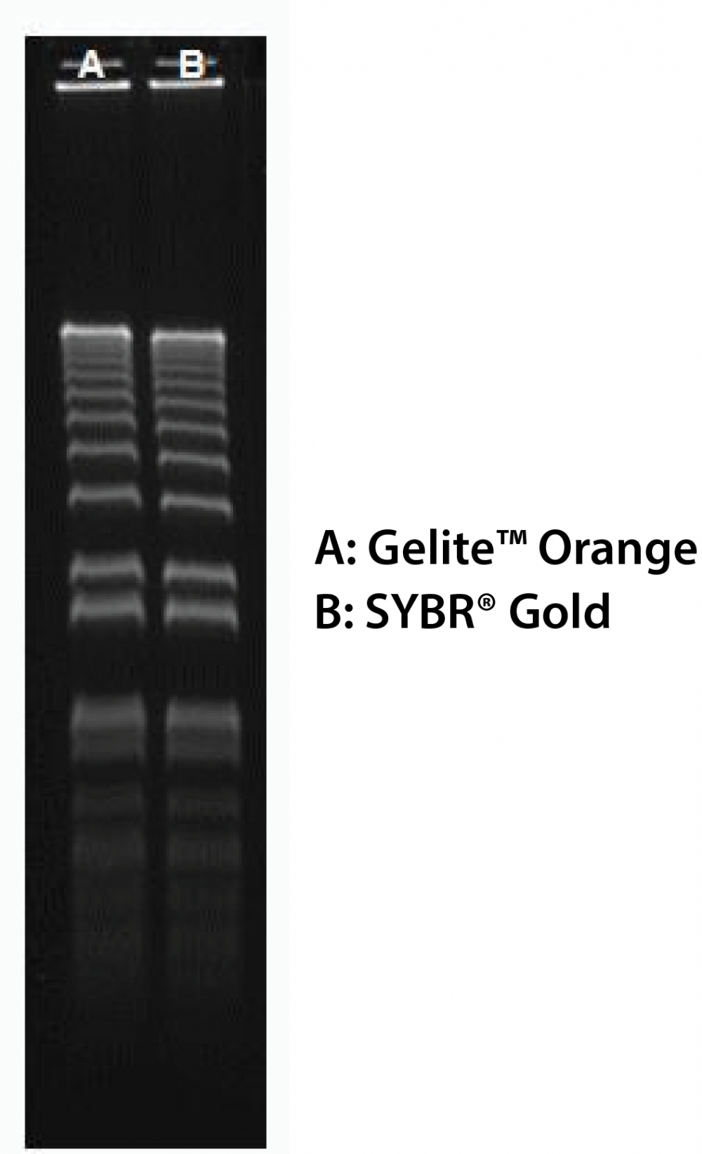
160 ng of 1 kb Plus DNA Ladder  (ThermoFisher 10787018) in 0.9% agarose/TBE electrophoresis gel were stained with Gelite™ Orange and SYBR® Gold, and imaged with 254-nm UV transilluminator using UVP Bioimaging System.
Ordering information
Price
Unit size
Catalog Number17594
Quantity
Add to cart
Additional ordering information
Telephone1-800-990-8053
Fax1-800-609-2943
Emailsales@aatbio.com
InternationalSee distributors
Bulk requestInquire
Custom sizeInquire
ShippingStandard overnight for United States, inquire for international
Request quotation
Storage, safety and handling
H-phraseH303, H313, H340
Hazard symbolT
Intended useResearch Use Only (RUO)
R-phraseR20, R21, R68
UNSPSC41116134
Platform

Transilluminator

Excitation254 nm or 300 nm
EmissionLong path green filter (ex. SYBR or GelStar)
Components
Example protocol

PREPARATION OF WORKING SOLUTION

Add 1 μL of Gelite™ Orange (Component A) into 200 μL of 5X Gel Loading Buffer (Component B). Protect the working solution from light by covering it with foil or placing it in the dark.

SAMPLE EXPERIMENTAL PROTOCOL

  1. Prepare DNA samples as you desired.
  2. Add 4 µL of Gelite™ Orange working solution into 16 µL of DNA samples and mix well. Incubate at room temperature for 5 - 15 minutes prior to electrophoresis.
  3. Run gels based on your standard protocol.
  4. Image the gel with a 300 nm ultraviolet or 254 nm transilluminator, or a laser-based gel scanner using a long path green filter such as a SYBR® filter or GelStar® filter. 
References
View all 79 references: Citation Explorer
SYBR green real-time PCR for the detection of all enterovirus-A71 genogroups
Authors: Dubot-Peres A, Tan CY, de Chesse R, Sibounheuang B, Vongsouvath M, Phommasone K, Bessaud M, Gazin C, Thirion L, Phetsouvanh R, Newton PN, de Lamballerie X.
Journal: PLoS One (2014): e89963
Development of loop-mediated isothermal amplification and SYBR green real-time PCR methods for the detection of Citrus yellow mosaic badnavirus in citrus species
Authors: Anthony Johnson AM, Dasgupta I, Sai Gopal DV.
Journal: J Virol Methods (2014): 9
Development of both multiple PCR and real-time SYBR green PCR for the detection of Vibrio cholerae non-O1/O139 serogroups
Authors: Li F, Kan B, Wang D.
Journal: Zhonghua Liu Xing Bing Xue Za Zhi (2014): 66
Detection of Cardamom mosaic virus and Banana bract mosaic virus in cardamom using SYBR Green based reverse transcription-quantitative PCR
Authors: Siljo A, Bhat AI, Biju CN.
Journal: Virusdisease (2014): 137
Comparison of SYBR Green and TaqMan methods in quantitative real-time polymerase chain reaction analysis of four adenosine receptor subtypes
Authors: Tajadini M, Panjehpour M, Javanmard SH.
Journal: Adv Biomed Res (2014): 85