logo
AAT Bioquest

Helixyte™ Green dsDNA Quantifying Reagent *200X DMSO Solution*

Comparison of calf thymus DNA dose response with Helixyte Green™ (blue circle) and PicoGreen® (red square) in a solid black 96-well microplate using a Gemini fluorescence microplate reader.
Ordering information
Price
Catalog Number
Unit Size
Quantity
Add to cart
Additional ordering information
Telephone1-800-990-8053
Fax1-800-609-2943
Emailsales@aatbio.com
InternationalSee distributors
Bulk requestInquire
Custom sizeInquire
ShippingStandard overnight for United States, inquire for international
Request quotation
Physical properties
Molecular weight661.17
SolventDMSO
Spectral properties
Excitation (nm)502
Emission (nm)522
Storage, safety and handling
H-phraseH303, H313, H340
Hazard symbolT
Intended useResearch Use Only (RUO)
R-phraseR20, R21, R68
StorageFreeze (< -15 °C); Minimize light exposure
UNSPSC41116134

OverviewpdfSDSpdfProtocol


Molecular weight
661.17
Excitation (nm)
502
Emission (nm)
522
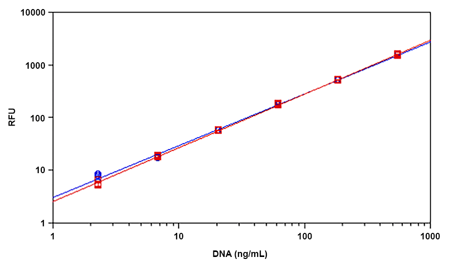
Helixyte™ Green is an excellent nucleic acid sensor that exhibits large fluorescence enhancement upon binding to dsDNA. It has the same spectral properties to those of PicoGreen®, thus a great replacement to PicoGreen® (PicoGreen® is the trademark of Invitrogen). The most commonly used technique for measuring nucleic acid concentration is the determination of absorbance at 260 nm (A260). However, the absorbance method suffers great interferences resulted from various contaminants commonly found in nucleic acid preparations, including nucleotides, single-stranded nucleic acids and proteins. Helixyte™ Green dsDNA Quantifying Reagent is an excellent alternative for quantifying DNAs with greatly improved sensitivity and selectivity. Helixyte™ Green dsDNA Quantifying Reagent is an ultra-sensitive fluorescent nucleic acid stain for quantitating double-stranded DNA (dsDNA) in molecular biological procedures such as cDNA synthesis for library production and DNA fragment purification for subcloning, as well as diagnostic applications, such as quantitating DNA amplification products and primer extension assays. Using the Helixyte™ Green dsDNA Quantifying Reagent, you can selectively detect as little as 25 pg/ml of dsDNA in the presence of ssDNA, RNA, and free nucleotides. The assay is linear over three orders of magnitude and has little sequence dependence, allowing you to accurately measure DNA from many sources, including genomic DNA, viral DNA, miniprep DNA, or PCR amplification products. Helixyte™ Green dsDNA Quantifying Reagent has a few orders of magnitude more sensitive than the UV absorbance readings, saving on precious sample. It is specific for dsDNA in the presence of equimolar amounts of RNA.

Platform


Fluorescence microplate reader

Excitation490
Emission525
Cutoff515
Recommended plateSolid black

Example protocol


PREPARATION OF WORKING SOLUTION

The following example is a protocol for quantifying dsDNA with Helixyte Green™. Before opening, allow the vial of Helixyte Green™ to warm to room temperature.


Note: Exercise caution when working with Helixyte Green™ dsDNA stain, as there is currently no available data regarding its potential mutagenicity or toxicity. Due to its affinity for nucleic acids, this reagent should be handled as a potential mutagen and with appropriate care. Additionally, take special care when handling the DMSO stock solution, as DMSO has been shown to facilitate the entry of organic molecules into tissues.

Preparing the Helixyte Green™ working solution
  1. To prepare an aqueous working solution of Helixyte Green™, dilute the concentrated DMSO solution 200-fold in TE buffer (10 mM Tris-HCl, 1 mM EDTA, pH 7.5-8.0). For example, adding 50 μL of Helixyte Green™ to 10 mL of TE will prepare enough working solution to assay 100 samples in a final volume of 200 µL. Protect the working solution from light by covering it with foil or placing it in the dark.
     
    Note: We recommend preparing this solution in a plastic container rather than glass, as the dye may adsorb to glass surfaces.
     
    Note:  For best results, this solution should be used within a few hours of its preparation. 

SAMPLE EXPERIMENTAL PROTOCOL

Prepare serial dilutions of dsDNA standard (0 to 3 ng/mL)
  1. Prepare a 1 mg/mL stock solution of dsDNA (such as calf thymus DNA from Sigma) in ddH2O

  2. To prepare a 10 μg/mL dsDNA solution, add 10 μL of the 1 mg/mL dsDNA stock solution (from Step 2.1) to 990 µL of TE buffer. Next, perform 1:10 and 1:2 serial dilutions to obtain concentrations of 1000, 100, 50, 25, 12.5, 6.25, 3.125, and 0 ng/mL.

  3. Add the dsDNA standards and test samples containing DNA into a solid black 96-well microplate, following the instructions provided in Tables 1 and 2.

Table 1. The layout of dsDNA standards and test samples in a solid black 96-well microplate.*

BL
BL

TS

TS

....

....


DS1

DS1

....

....

....

....


DS2

DS2

....

....

....

....


DS3

DS3


DS4

DS4


DS5

DS5


DS6

DS6


DS7

DS7


*Note: DS = dsDNA Standards; BL= Blank Control; TS =Test Samples

Table 2. Reagent composition for each well.* 

dsDNA Standard

Blank Control

Test Sample

Serial dilutions* (100 µL)

TE: 100 μL

100 μL

*Note: Add the serial dilutions of dsDNA standards from 0.1 to 1000 ng/mL into wells from DS1 to DS7 in duplicate. 

Run dsDNA assay
  1. Add 100 μL of dsDNA assay mixture (from Step 1.1) to each well of the dsDNA standard, blank control, and test samples (see Step 2.3) to make the total dsDNA assay volume of 200 µL/well.

    Note: For a 384-well plate, add 25 μL sample and 25 μL of dsDNA assay mixture per well.

    Note: For cuvette-based assays, add 1mL sample and 1mL of dsDNA assay mixture per cuvette.

  2. Incubate the reaction at room temperature for 5 to 10 minutes, protected from light.

  3. Monitor the fluorescence increase with a fluorescence microplate reader at Ex/Em = 490/525 nm, cut off at 515 nm.

  4. The fluorescence obtained in the blank wells (containing only TE buffer) is used as a control and will be subtracted from the values obtained in the wells containing dsDNA reactions. The DNA concentrations of the test samples can be determined by referencing the standard curve generated in the DNA Standard Curve step.

Calculators


Common stock solution preparation

Table 1. Volume of DMSO needed to reconstitute specific mass of Helixyte™ Green dsDNA Quantifying Reagent *200X DMSO Solution* to given concentration. Note that volume is only for preparing stock solution. Refer to sample experimental protocol for appropriate experimental/physiological buffers.

0.1 mg0.5 mg1 mg5 mg10 mg
1 mM151.247 µL756.235 µL1.512 mL7.562 mL15.125 mL
5 mM30.249 µL151.247 µL302.494 µL1.512 mL3.025 mL
10 mM15.125 µL75.624 µL151.247 µL756.235 µL1.512 mL

Molarity calculator

Enter any two values (mass, volume, concentration) to calculate the third.

Mass (Calculate)Molecular weightVolume (Calculate)Concentration (Calculate)Moles
/=x=

Spectrum


Open in Advanced Spectrum Viewer
spectrum

Spectral properties

Excitation (nm)502
Emission (nm)522

Images


Citations


View all 4 citations: Citation Explorer
Bovine Fibroblast-Derived Extracellular Matrix Promotes the Growth and Preserves the Stemness of Bovine Stromal Cells during In Vitro Expansion
Authors: Lee, Kathleen and Jackson, Anisha and John, Nikita and Zhang, Ryan and Ozhava, Derya and Bhatia, Mohit and Mao, Yong
Journal: Journal of Functional Biomaterials (2023): 218
Arthropods, nematodes, fungi, and bacteria associated with penguin carrion in Barton Peninsula, King George Island, Antarctica
Authors: ZAINI, NUR ADILLA and Low, Van Lun and Gebrelassie, Selam Solomon and Ismail, Siti Sofo and Mahmud, Mohd Hafizi and Houssaini, Jamal and Lee, Won Young and Tomberlin, Jeffery K and Heo, Chong Chin
Journal: (2023)
Faecal DNA metabarcoding reveals novel bacterial community patterns of critically endangered Southern River Terrapin, Batagur affinis
Authors: Salleh, Mohd Hairul Mohd and Esa, Yuzine and Ngalimat, Mohamad Syazwan and Chen, Pelf Nyok
Journal: PeerJ (2022): e12970
Archaeal and methanogenic communities in the rice field under different fertilizer applications
Authors: Fatma, Yuli and RUSMANA, IMAN and WAHYUDI, ARIS TRI and others,
Journal: Biodiversitas Journal of Biological Diversity (2019)

References


View all 31 references: Citation Explorer
Inhibitors of Streptococcus pneumoniae surface endonuclease EndA discovered by high-throughput screening using a PicoGreen fluorescence assay
Authors: Peterson EJ, Kireev D, Moon AF, Midon M, Janzen WP, Pingoud A, Pedersen LC, Singleton SF.
Journal: J Biomol Screen (2013): 247
Validation of a PicoGreen-based DNA quantification integrated in an RNA extraction method for two-dimensional and three-dimensional cell cultures
Authors: Chen Y, Sonnaert M, Roberts SJ, Luyten FP, Schrooten J.
Journal: Tissue Eng Part C Methods (2012): 444
Characterization of PicoGreen interaction with dsDNA and the origin of its fluorescence enhancement upon binding
Authors: Dragan AI, Casas-Finet JR, Bishop ES, Strouse RJ, Schenerman MA, Geddes CD.
Journal: Biophys J (2010): 3010
Comparison of SYBR Green I-, PicoGreen-, and [3H]-hypoxanthine-based assays for in vitro antimalarial screening of plants from Nigerian ethnomedicine
Authors: Abiodun OO, Gbotosho GO, Ajaiyeoba EO, Happi CT, Hofer S, Wittlin S, Sowunmi A, Brun R, Oduola AM.
Journal: Parasitol Res (2010): 933
Metal-enhanced PicoGreen fluorescence: application to fast and ultra-sensitive pg/ml DNA quantitation
Authors: Dragan AI, Bishop ES, Casas-Finet JR, Strouse RJ, Schenerman MA, Geddes CD.
Journal: J Immunol Methods (2010): 95
Quantification of dsDNA using the Hitachi F-7000 Fluorescence Spectrophotometer and PicoGreen dye
Authors: Moreno LA, Cox KL.
Journal: J Vis Exp. (2010)
Factors affecting quantification of total DNA by UV spectroscopy and PicoGreen fluorescence
Authors: Holden MJ, Haynes RJ, Rabb SA, Satija N, Yang K, Blasic JR, Jr.
Journal: J Agric Food Chem (2009): 7221
Development and characterization of a novel host cell DNA assay using ultra-sensitive fluorescent nucleic acid stain "PicoGreen"
Authors: Ikeda Y, Iwakiri S, Yoshimori T.
Journal: J Pharm Biomed Anal (2009): 997
Enhanced DNA dynamics due to cationic reagents, topological states of dsDNA and high mobility group box 1 as probed by PicoGreen
Authors: Noothi SK, Kombrabail M, Kundu TK, Krishnamoorthy G, Rao BJ.
Journal: FEBS J (2009): 541
Label-free DNA sequence detection with enhanced sensitivity and selectivity using cationic conjugated polymers and PicoGreen
Authors: Ren X, Xu QH.
Journal: Langmuir (2009): 43