logo
AAT Bioquest

Phalloidin-iFluor® 488 Conjugate

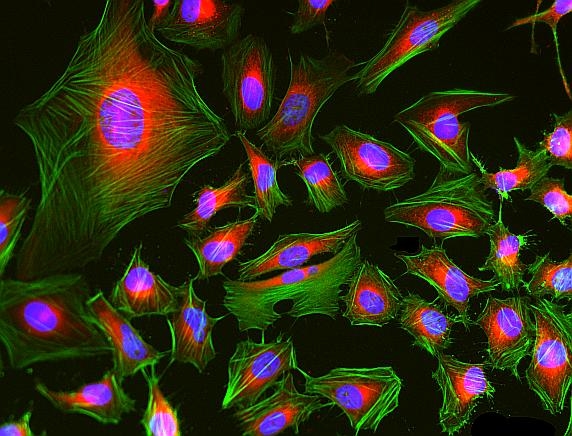
Fluorescence images of HeLa cells stained with Phalloidin-iFluor® 488 Conjugate using fluorescence microscope with a FITC filter set (Green). The cells were fixed in 4% formaldehyde, co-labeled with mitochondria dye MitoLite™ Red FX600 (Cat#2677, Red) and Nuclear Blue™ DCS1 (Cat#17548, Blue).
<strong>Figure 2.</strong> MDA-MB-231 breast cancer cell grew for 24 h. Cells were stained with Phalloidin-iFluor 488 Conjugate (ATT Bioquest) following manufacturer&rsquo;s instruction. Images were acquired with a 63x/1.4NA objective on a Zeiss laser-scanning confocal microscope by the Advanced Bio-Imaging Facility (ABIF) at McGill.&nbsp;Displayed is the Max Intensity Projection of 19 images with 0.2 um spacing in Z.
Enterobacteriaceae (Lipid A) in the liver of the uninfected and the liver fluke-infected hamsters. a. An uninfected hamster. b. 3D reconstruction of the internal surface of a bile duct after confocal microscopy reveals the presence of Enterobacteriaceae Lipid A. c. Bacteria inside the gut of the O. viverrini parasite. d. Enterobacteriaceae presence inside small bile ducts of an O. viverrini–infected hamster. e. Penetration of bacteria through the injured epithelium in the bile duct of an O. felineus–infected hamster. f. A multilayered epithelium in the bile duct of a C. sinensis–infected hamster. E: epithelial cells; BD: bile duct; red color: Lipid A of Enterobacteriaceae; green color: actin filaments (Phalloidin 488 staining); blue color: nuclei (DAPI staining). E: epithelium of bile duct; BD: bile duct; G: gut of a worm. Source: <b><em>Opisthorchis viverrini</em>, <em>Clonorchis sinensis</em> and <em>Opisthorchis felineus</em> liver flukes affect mammalian host microbiome in a species-specific manner</b> by Pakharukova <em>et. al.</em>, <em>PLoS Negl Trop Dis</em>. Feb. 2023.
Conditioning of GelMA-AlgMA bioinks for skeletal muscle tissue engineering. Modulation of GelMA-AlgMA bioink mechanical properties of GelMA with 0 %,1 % and 2 % AlgMA (n = 9). Confocal images of C2C12 cells after 14 days of differentiation with stained MHC (red), F-actin (green) and nuclei (blue). Actin was stained with Phalloidin-iFluor 488. Scale bar = 100 μm. Source: <b>3D bioprinted functional skeletal muscle models have potential applications for studies of muscle wasting in cancer cachexia</b> by Andrea García-Lizarribar et.al., <em>Biomaterials Advances</em> April 2023.
Myogenic differentiation in 3D bioprinted models. Immunostaining of C2C12 cells after 15 days in bioprinted rings cultured in differentiation medium (DM) and growth medium (GM). Nuclei are stained in blue and green corresponds to F-actin (n = 4). Scale bar = 200 μm. Actin was stained with Phalloidin-iFluor 488. 
Source: <b>3D bioprinted functional skeletal muscle models have potential applications for studies of muscle wasting in cancer cachexia</b> by Andrea García-Lizarribar et.al., <em>Biomaterials Advances</em> April 2023.
Ordering information
Price
Catalog Number
Unit Size
Quantity
Add to cart
Additional ordering information
Telephone1-800-990-8053
Fax1-800-609-2943
Emailsales@aatbio.com
InternationalSee distributors
Bulk requestInquire
Custom sizeInquire
ShippingStandard overnight for United States, inquire for international
Request quotation
Physical properties
Molecular weight~1400
SolventDMSO
Spectral properties
Correction Factor (260 nm)0.21
Correction Factor (280 nm)0.11
Extinction coefficient (cm -1 M -1)750001
Excitation (nm)491
Emission (nm)516
Quantum yield0.91
Storage, safety and handling
Certificate of OriginDownload PDF
H-phraseH301, H311, H331
Hazard symbolT
Intended useResearch Use Only (RUO)
R-phraseR23, R24, R25
StorageFreeze (< -15 °C); Minimize light exposure
UNSPSC12352200

OverviewpdfSDSpdfProtocol


Molecular weight
~1400
Correction Factor (260 nm)
0.21
Correction Factor (280 nm)
0.11
Extinction coefficient (cm -1 M -1)
750001
Excitation (nm)
491
Emission (nm)
516
Quantum yield
0.91
This green fluorescent phalloidin conjugate (equivalent to Alexa Fluor® 488-labeled phalloidin) selectively binds to F-actins with much higher photostability than the fluorescein-phalloidin conjugates. Used at nanomolar concentrations, phalloidin derivatives are convenient probes for labeling, identifying and quantitating F-actins in formaldehyde-fixed and permeabilized tissue sections, cell cultures or cell-free experiments. Phalloidin binds to actin filaments much more tightly than to actin monomers, leading to a decrease in the rate constant for the dissociation of actin subunits from filament ends, essentially stabilizing actin filaments through the prevention of filament depolymerization. Moreover, phalloidin is found to inhibit the ATP hydrolysis activity of F-actin. Phalloidin functions differently at various concentrations in cells. When introduced into the cytoplasm at low concentrations, phalloidin recruits the less polymerized forms of cytoplasmic actin as well as filamin into stable "islands" of aggregated actin polymers, yet it does not interfere with stress fibers, i.e. thick bundles of microfilaments. The property of phalloidin is a useful tool for investigating the distribution of F-actin in cells by labeling phalloidin with fluorescent analogs and using them to stain actin filaments for light microscopy. Fluorescent derivatives of phalloidin have turned out to be enormously useful in localizing actin filaments in living or fixed cells as well as for visualizing individual actin filaments in vitro. Fluorescent phalloidin derivatives have been used as an important tool in the study of actin networks at high resolution. AAT Bioquest offers a variety of fluorescent phalloidin derivatives with different colors for multicolor imaging applications.

Example protocol


AT A GLANCE

Protocol Summary
  1. Prepare samples in microplate wells
  2. Remove liquid from samples in the plate
  3. Add Phalloidin-iFluor™ 488 Conjugate solution (100 μL/well)
  4. Stain the cells at room temperature for 20 to 90 minutes
  5. Wash the cells
  6. Examine the specimen under microscope with FITC filter 
Important      Warm the vial to room temperature and centrifuge briefly before opening.

Storage and Handling Conditions
The solution should be stable for at least 6 months if store at -20 °C. Protect the fluorescent conjugates from light, and avoid freeze/thaw cycles.
Note     Phalloidin is toxic, although the amount of toxin present in a vial could be lethal only to a mosquito (LD50 of phalloidin = 2 mg/kg), it should be handled with care.

PREPARATION OF WORKING SOLUTION

Phalloidin-iFluor™ 488 Conjugate working solution
Add 1 µL of Phalloidin-iFluor™ 488 Conjugate solution to 1 mL of PBS with 1% BSA.
Note     The stock solution of phalloidin conjugate should be aliquoted and stored at -20 °C. protected from light.
Note     Different cell types might be stained differently. The concentration of phalloidin conjugate working solution should be prepared accordingly.

SAMPLE EXPERIMENTAL PROTOCOL

Stain the cells
  1. Perform formaldehyde fixation. Incubate cells with 3.0–4.0 % formaldehyde in PBS at room temperature for 10–30 minutes.
    Note     Avoid any methanol containing fixatives since methanol can disrupt actin during the fixation process. The preferred fixative is methanol-free formaldehyde.
  2. Rinse the fixed cells 2–3 times in PBS.
  3. Optional: Add 0.1% Triton X-100 in PBS into fixed cells for 3 to 5 minutes to increase permeability. Rinse the cells 2–3 times in PBS.
  4. Add 100 μL/well (96-well plate) of Phalloidin-iFluor™ 488 Conjugate working solution into the fixed cells, and stain the cells at room temperature for 20 to 90 minutes.
  5. Rinse cells gently with PBS 2 to 3 times to remove excess phalloidin conjugate before plating, sealing and imaging under microscope with FITC filter set. 

Spectrum


Open in Advanced Spectrum Viewer
spectrum

Spectral properties

Correction Factor (260 nm)0.21
Correction Factor (280 nm)0.11
Extinction coefficient (cm -1 M -1)750001
Excitation (nm)491
Emission (nm)516
Quantum yield0.91

Product Family


NameExcitation (nm)Emission (nm)Extinction coefficient (cm -1 M -1)Quantum yieldCorrection Factor (260 nm)Correction Factor (280 nm)
Phalloidin-iFluor® 350 Conjugate3454502000010.9510.830.23
Phalloidin-iFluor® 405 Conjugate4034273700010.9110.480.77
Phalloidin-iFluor® 514 Conjugate5115277500010.8310.2650.116
Phalloidin-iFluor® 532 Conjugate5375609000010.6810.260.16
Phalloidin-iFluor® 555 Conjugate55757010000010.6410.230.14
Phalloidin-iFluor® 594 Conjugate58760320000010.5310.050.04
Phalloidin-iFluor® 633 Conjugate64065425000010.2910.0620.044
Phalloidin-iFluor® 647 Conjugate65667025000010.2510.030.03
Phalloidin-iFluor® 680 Conjugate68470122000010.2310.0970.094
Phalloidin-iFluor® 700 Conjugate69071322000010.2310.090.04
Phalloidin-iFluor® 750 Conjugate75777927500010.1210.0440.039
Phalloidin-iFluor® 790 Conjugate78781225000010.1310.10.09
iFluor® 488-streptavidin conjugate4915167500010.910.210.11
Show More (4)

Images


Citations


View all 311 citations: Citation Explorer
NIR-activated electrospun nanodetonator dressing enhances infected diabetic wound healing with combined photothermal and nitric oxide-based gas therapy
Authors: Xie, Jiajun and Liu, Guihua and Chen, Rong and Wang, Ding and Mai, Huaming and Zhong, Qiang and Ning, Yanhong and Fu, Jinlang and Tang, Zinan and Xu, Yixin and others,
Journal: Journal of Nanobiotechnology (2024): 1--25
SOX4 induces cytoskeleton remodeling and promotes cell motility via N-WASP/ARP2/3 pathway in colorectal cancer cells
Authors: Anupriya, S and Parida, Nandita and Patnaik, Srinivas
Journal: Experimental Cell Research (2024): 114059
3D in vitro synovial hyperplasia model on polycaprolactone-micropatterned nanofibrous microwells for screening disease-modifying anti-rheumatic drugs
Authors: Kim, Dongwoo and Heo, Jiyeon and Song, Boa and Lee, Gyubok and Hong, Changgi and Jiang, Zhuomin and Lee, Sohui and Lee, Kangwon and Kim, Mingyo and Park, Min Hee
Journal: Materials Today Bio (2024): 101061
Physiological DNA damage promotes functional endoreplication of mammary gland alveolar cells during lactation
Authors: Molinuevo, Rut and Menendez, Julien and Cadle, Kora and Ariqat, Nabeela and Choy, Marie Klaire and Lagousis, Cayla and Thomas, Gwen and Strietzel, Catherine and Bubolz, JW and Hinck, Lindsay
Journal: Nature Communications (2024): 3288
The BKCa (slo) channel regulates the cardiac function of Drosophila
Authors: Gururaja Rao, Shubha and Lam, Alexander and Seeley, Sarah and Park, Jeniffer and Aruva, Shriya and Singh, Harpreet
Journal: Physiological Reports (2024): e15996
Chronic hypoxia remodels the tumor microenvironment to support glioma stem cell growth
Authors: Nicholson, JG and Cirigliano, S and Singhania, R and Haywood, C and Shahidi Dadras, M and Yoshimura, M and Vanderbilt, D and Liechty, B and Fine, HA
Journal: Acta Neuropathologica Communications (2024): 46
The exon junction complex is required for DMD gene splicing fidelity and myogenic differentiation
Authors: Da Cunha, Dylan and Miro, Julie and Van Goethem, Charles and Notarnicola, C{\'e}cile and Hugon, G{\'e}rald and Carnac, Gilles and Coss{\'e}e, Mireille and Koenig, Michel and Tuffery-Giraud, Sylvie
Journal: Cellular and Molecular Life Sciences (2024): 150
Quantitative Investigation of Intestinal Drug Absorption Enhancement by Drug-Rich Nanodroplets Generated via Liquid--Liquid Phase Separation
Authors: Yoshikawa, Etsushi and Ueda, Keisuke and Hakata, Rei and Higashi, Kenjirou and Moribe, Kunikazu
Journal: Molecular Pharmaceutics (2024)
Micro-alloying of Zn and Ca in Vacuum Induction Casted Bioresorbable Mg System: Perspectives on Corrosion Resistance, Cytocompatibility, and Inflammatory Response
Authors: Behera, Manisha and Denys, Agnes and Shabadi, Rajashekhara and Allain, Fabrice and Gruescu, Cosmin
Journal: (2024)

References


View all 127 references: Citation Explorer
Phalloidin perturbs the interaction of human non-muscle myosin isoforms 2A and 2C1 with F-actin
Authors: Diensthuber RP, Muller M, Heissler SM, Taft MH, Chizhov I, Manstein DJ.
Journal: FEBS Lett (2011): 767
Improved penile histology by phalloidin stain: circular and longitudinal cavernous smooth muscles, dual-endothelium arteries, and erectile dysfunction-associated changes
Authors: Lin G, Qiu X, F and el TM, Albersen M, Wang Z, Lue TF, Lin CS.
Journal: Urology (2011): 970 e1
Labeling cytoskeletal F-actin with rhodamine phalloidin or fluorescein phalloidin for imaging
Authors: Chazotte B., undefined
Journal: Cold Spring Harb Protoc (2010): pdb prot4947
pH-(low)-insertion-peptide (pHLIP) translocation of membrane impermeable phalloidin toxin inhibits cancer cell proliferation
Authors: An M, Wijesinghe D, Andreev OA, Reshetnyak YK, Engelman DM.
Journal: Proc Natl Acad Sci U S A (2010): 20246
Pygmy squids and giant brains: mapping the complex cephalopod CNS by phalloidin staining of vibratome sections and whole-mount preparations
Authors: Wollesen T, Loesel R, Wanninger A.
Journal: J Neurosci Methods (2009): 63
Processing of the phalloidin proprotein by prolyl oligopeptidase from the mushroom Conocybe albipes
Authors: Luo H, Hallen-Adams HE, Walton JD.
Journal: J Biol Chem (2009): 18070
In vitro inhibition of OATP-mediated uptake of phalloidin using bile acid derivatives
Authors: Herraez E, Macias RI, Vazquez-Tato J, Vicens M, Monte MJ, Marin JJ.
Journal: Toxicol Appl Pharmacol (2009): 13
Effect of Phalloidin on Filaments Polymerized from Heart Muscle Adp-Actin Monomers
Authors: Vig A, Dudas R, Kupi T, Orban J, Hild G, Lorinczy D, Nyitrai M.
Journal: J Therm Anal Calorim (2009): 721
Protective effect of bile acid derivatives in phalloidin-induced rat liver toxicity
Authors: Herraez E, Macias RI, Vazquez-Tato J, Hierro C, Monte MJ, Marin JJ.
Journal: Toxicol Appl Pharmacol (2009): 21
Anti-acetylated tubulin antibody staining and phalloidin staining in the starlet sea anemone Nematostella vectensis
Authors: Genikhovich G, Technau U.
Journal: Cold Spring Harb Protoc (2009): pdb prot5283