Products
Services
Resources
Selection Guides
About
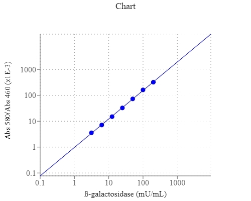
Amplite® Colorimetric Beta-Galactosidase Assay Kit
E. coli beta-galactosidase is a 464 kD tetramer. Each unit of beta-galactosidase consists of five domains, the third of which is the active site. It is an essential enzyme in cells. Deficiencies of this enzyme can result in galactosialidosis or Morquio B syndrome. In E. coli, beta-galactosidase is produced by the activation of LacZ operon. Detection of LacZ expression has become routine to the point of detection of as few as 5 copies of beta-galactosidase per cell. This kit uses a chromogenic galactosidase substrate that can sensitively distinguish LacZ+ from LacZ- cells. The light yellow substrate generates a strongly purple product upon reaction with galactosidase. It can be used either for detecting galactosidase conjugates in ELISA type assay systems or for monitoring LacZ gene expression in cells. Amplite® Colorimetric Beta-Galactosidase Assay Kit comes with all the essential components with an optimized assay protocol. It can be used for screening galactosidase inhibitors or inducers.
| Catalog | Size | Price | Quantity |
|---|---|---|---|
| 12604 | 200 Tests | Price |
Usage and storage
| Intended use | Research Use Only (RUO) |
Instrument settings
| Absorbance microplate reader | |
| Absorbance | 580/460 nm |
| Recommended plate | Clear bottom |
Contact us
| Telephone | |
| Fax | |
| sales@aatbio.com | |
| International | See distributors |
| Bulk request | Inquire |
| Custom size | Inquire |
| Technical Support | Contact us |
| Request quotation | Request |
| Purchase order | Send to sales@aatbio.com |
| Shipping | Standard overnight for United States, inquire for international |
Page updated on July 17, 2026