logo
AAT Bioquest

Amplite® Fluorimetric HDAC Activity Assay Kit *Green Fluorescence*

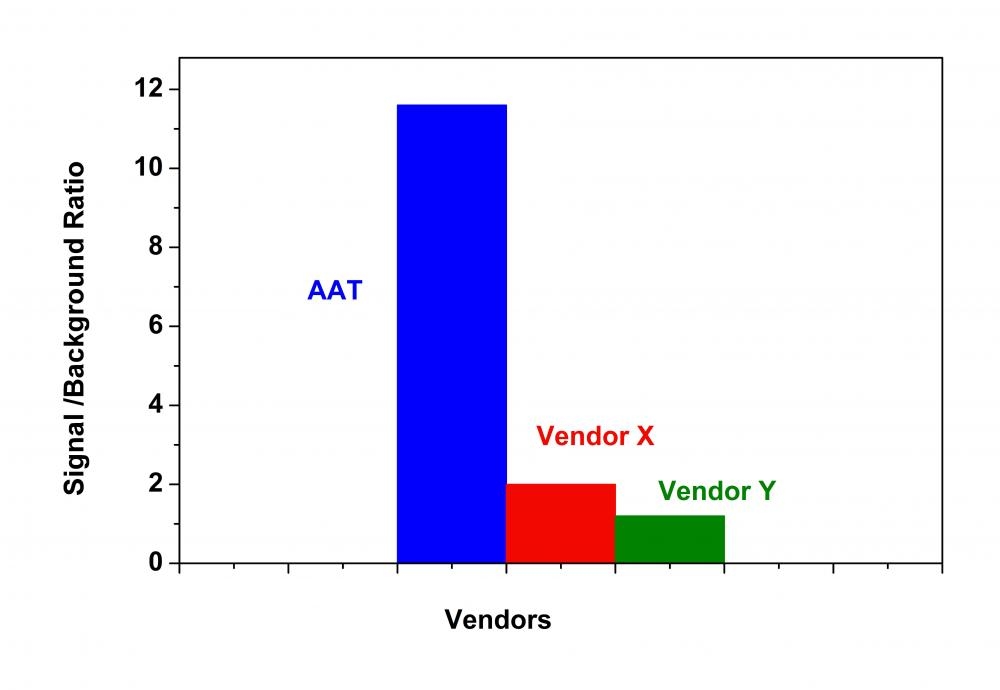
HDAC activity in HeLa nuclear extract was measured with Amplite® Fluorimetric HDAC Activity Assay Kit (in blue) was compared with Vendor X (in red) and Vendor Y (in green), both of which use Ac-RGK(Ac)-R110 peptide substrate. The signal/background ratio of the HDAC activity measured with Amplite® Fluorimetric HDAC Activity Assay Kit is more than 10 times higher than those of Vendors X and Y.
Trichostatin A inhibition in HeLa nuclear extract was measured with Amplite® Fluorimetric HDAC Activity Assay Kit using Gemini Fluorescence microplate reader (Molecular Devices).
Butyrate upregulates HDP expression via HDAC inhibition. (A) Overall inhibition of HDAC enzymes in 3D4/2 cell nuclear extracts by increasing concentrations of butyrate. TSA was used as a reference. (B) HDAC activity in 3D4/2 cells was determined. Cells were incubated with NaB, TSA or an HAT inhibitor for 24h. The results were normalized using the control as 100%. ***Indicate an extreme significant difference (p&thinsp;&lt;&thinsp;0.001) compared to that of control group by One-way ANOVA. Western blot (C) and immunofluorescence (D) analysis of acetylated histone complex H3 in 3D4/2 cells. 3D4/2 cells were incubated with NaB, TSA or HAT inhibitor for 24&thinsp;h. Western blot analysis of the acetylation of H3 (AcH3) was examined in the nuclear extract. Immunofluorescent staining with anti-AcH3 (red) Ab. Cells were counterstained with DAPI. Effects of butyrate, TSA and HAT inhibitor on HDP gene expression in macrophage 3D4/2 cells. (E) 3D4/2 cells were treated with NaB, TSA and an HAT inhibitor for 24&thinsp;h. cDNA was analyzed by qPCR. Gene expression changes are expressed as the foldchange in transcription in treatedcells with respect to un-treated cells. Numbers &lt;1 denote downregulation, and those &gt;1 indicate upregulation. Data were normalized to GAPDH. (F) 3D4/2 cells were treated with NaB, TSA or an HAT inhibitor for 24&thinsp;h. Samples were analyzed by ChIP using antibodies against H3K9Ac. Purified DNA was analyzed by qPCR using primers specific to the promoters of the indicated genes. Normalized results are&nbsp;shown as a percentage of the input values. Source: <strong>Butyrate upregulates endogenous host defense peptides to enhance disease resistance in piglets via histone deacetylase inhibition </strong>by Xiong et al., <em>Scientific Reports</em>, May 2016.
Ordering information
Price
Catalog Number
Unit Size
Quantity
Add to cart
Additional ordering information
Telephone1-800-990-8053
Fax1-800-609-2943
Emailsales@aatbio.com
InternationalSee distributors
Bulk requestInquire
Custom sizeInquire
ShippingStandard overnight for United States, inquire for international
Request quotation
Spectral properties
Excitation (nm)493
Emission (nm)520
Storage, safety and handling
Certificate of OriginDownload PDF
H-phraseH303, H313, H333
Hazard symbolXN
Intended useResearch Use Only (RUO)
R-phraseR20, R21, R22
UNSPSC12352200

OverviewpdfSDSpdfProtocol


Excitation (nm)
493
Emission (nm)
520
Histone deacetylases (HDAC) are a class of enzymes that remove acetyl groups from a ε-N-acetyl lysine amino acid on a histone. Deacetylation restores the positive electric charge of the lysine amino acids, which increases the histone's affinity for the negatively charged phosphate backbone of DNA. This generally down-regulates DNA transcription by blocking the access of transcription factors. HDACs are involved in the pathway by which the retinoblastoma protein (pRb) suppresses cell proliferation. The pRb protein is part of a complex which attracts HDACs to the chromatin so that it will deacetylate histones. HDAC inhibitors are being studied as a treatment for cancer. The Amplite® HDAC Assay Kit provides a quick, convenient, and sensitive method for the detection of HDAC activity. Our HDAC Green™ substrate is a non-peptide compound that is much more resistant than other commercial peptide-based HDAC substrates. Our kit can be used for measuring HDAC activity in cell lysates, in vitro inhibitor screening with extracts or purified enzymes. The long wavelength emission and higher extinction coefficient of the HDAC Green™ substrate provide less interference from compounds and cell components. HDAC activity is determined by monitoring the green fluorescence enhancement with excitation at 490 nm and emission at 520 nm.
Platform


Fluorescence microplate reader

Excitation490 nm
Emission525 nm
Cutoff515 nm
Recommended plateSolid black

Components


Example protocol


AT A GLANCE

Protocol Summary
  1. Prepare HDAC containing samples (40 µL)
  2. Add HDAC inhibitor or test compounds (10 µL)
  3. Incubate at room temperature or 37°C for 10 - 20 minutes
  4. Add HDAC Green™ Substrate working solution (50 µL)
  5. Incubate at room temperature or 37°C for 30 - 60 minutes
  6. Monitor fluorescence intensity at Ex/Em = 490/525 nm (Cutoff = 515 nm) 
Important      Thaw all the kit components before starting the experiment.

PREPARATION OF WORKING SOLUTION

1. HDAC-containing test samples
Dilute 5 - 10 mg/mL of HeLa nuclear extract or cell lysates at 1:40 in Assay Buffer (Component B). 
Note     40 µL of the diluted sample is enough for one well of a 96-well plate. Dilute extract immediately before use. Store the solution on ice.


2. HDAC inhibitor (Trichostain A) solution (30 µM)
Dilute 3 mM Trichostatin A solution (Component C) at 1:100 in assay buffer (Component B) to get a 30 µM Trichostatin A solution. Add 10 μL of the 30 μM Trichostatin A solution into each inhibitor control well.

3. HDAC Green™ Substrate working solution
Add 20 μL of HDAC Green™ Substrate (Component A) and 100 μL of the Signal Enhancer (Component D) into 5 mL of Assay Buffer (Component B). 
Note     The diluted HDAC Green™ Substrate working solution is not stable. 5 mL of the diluted HDAC Green™ Substrate working solution is enough for 100 assays. Prepare fresh HDAC Green™ Substrate working solution for each experiment. Keep reconstituted working solution on ice until use.

SAMPLE EXPERIMENTAL PROTOCOL

Table 1. Layout of nuclear extracts with test compounds in a solid black 96-well microplate.
SamplesHeLa ExtractAssay Buffer (Component B)Trichostatin A (HDAC inhibitor)Test CompoundsHDAC Green™ Substrate
Blank (no enzyme)0 µL50 µL0 µL0 µL50 µL
Positive Control40 µL10 µL0 µL0 µL50 µL
Negative Control40 µL0 µL10 µL0 µL50 µL
Test Compound40 µL0 µL0 µL10 µL50 µL
  1. Add 40 µL of diluted nuclear extract, enzyme solution or other HDAC samples, and 10 µL of test compounds to the corresponding microplate wells.
    1. For positive control: Add 40 µL of diluted HDAC enzyme solution or HeLa nuclear extract with 10 µL of Assay Buffer (Component B).
    2. For negative control: Add 40 µL of diluted HeLa nuclear extract with 10 µL of 30 µM Trichostatin A solution, or use a known sample containing no HDAC activity.
    3. For Blank (no Enzyme): Add 50 µL of Assay Buffer (Component B) only.
  2. Incubate the plate at room temperature or 37°C for 10 - 20 minutes.
    Note     For screening HDAC inhibitor, preincubate the compounds with HeLa nuclear extract or pure enzyme before adding HDAC Green™ Substrate working solution.
  3. Add 50 µL of HDAC Green™ Substrate working solution into each well. Incubate the plate at room temperature or 37°C for 30 - 60 minutes.
  4. Monitor fluorescence intensity at Ex/Em = 490/525 nm. (Cutoff = 515 nm) 

Spectrum


Open in Advanced Spectrum Viewer
spectrum

Spectral properties

Excitation (nm)493
Emission (nm)520

Images


Citations


View all 18 citations: Citation Explorer
Target-based drug discovery: Applications of fluorescence techniques in high throughput and fragment-based screening
Authors: Kumar, Vikrant and Lakshman, Puneeth Kumar Chunchagatta and Prasad, Thazhe Kootteri and Manjunath, Kavyashree and Bairy, Sneha and Vasu, Akshaya S and Ganavi, B and Jasti, Subbarao and Kamariah, Neelagandan
Journal: Heliyon (2023): e23864
Influence of Dinitrosyl Iron Complexes on the Activity of Enzymes, Indicators of Cardiovascular Diseases
Authors: Akentieva, Natalia and Sanina, Natalia and Gizatullin, Artur and Shkondina, Natalia and Andreeva, Anna and Shram, Stanislav and Aldoshin, Sergei
Journal: (2022)
Porcine deltacoronavirus infection cleaves HDAC2 to attenuate its antiviral activity
Authors: Li, Zhuang and Fang, Puxian and Duan, Panpan and Chen, Jiyao and Fang, Liurong and Xiao, Shaobo
Journal: Journal of Virology (2022): e01027--22
Histone deacetylase 5 deacetylates the phosphatase PP2A for positively regulating NF-$\kappa$B signaling
Authors: Xu, Chonghui and Tang, Jielin and Yang, Qi and Zhao, He and Liu, Yaling and Cao, Juan and Zhou, Yuan and Chen, Xinwen and Chen, Jizheng
Journal: Journal of Biological Chemistry (2021): 101380
Intestinal microbiota-derived short-chain fatty acids regulation of immune cell IL-22 production and gut immunity
Authors: Yang, Wenjing and Yu, Tianming and Huang, Xiangsheng and Bilotta, Anthony J and Xu, Leiqi and Lu, Yao and Sun, Jiaren and Pan, Fan and Zhou, Jia and Zhang, Wenbo and others,
Journal: Nature communications (2020): 1--18
Microbiota metabolite short-chain fatty acids facilitate mucosal adjuvant activity of cholera toxin through GPR43
Authors: Yang, Wenjing and Xiao, Yi and Huang, Xiangsheng and Chen, Feidi and Sun, Mingming and Bilotta, Anthony J and Xu, Leiqi and Lu, Yao and Yao, Suxia and Zhao, Qihong and others,
Journal: The Journal of Immunology (2019): 282--292
TLR2/EGFR are two Sensors for pBD3 and pEP2C induction by Sodium Butyrate independent on HDAC inhibition
Authors: Dou, Xiujing and Gao, Nan and Lan, Jing and Han, Junlan and Yang, Yang and Shan, Anshan
Journal: Journal of Agricultural and Food Chemistry (2019)
Minor structural modifications to Pracinostat produce big changes in its biological responses
Authors: Jia, Rong and Sun, Pengju and Zhang, Yan and Ge, Youjin and Yu, Niefang
Journal: Chemical biology \& drug design (2019): 1488--1493
NOS1 inhibits the interferon response of cancer cells by S-nitrosylation of HDAC2
Authors: Xu, Pengfei and Ye, Shuangyan and Li, Keyi and Huang, Mengqiu and Wang, Qianli and Zeng, Sisi and Chen, Xi and Gao, Wenwen and Chen, Jianping and Zhang, Qianbing and others, undefined
Journal: Journal of Experimental &amp; Clinical Cancer Research (2019): 1--16
Characterization of the Host Tissue Response Induced by a Biosynthetic Material Composed of Poly (4-Hydroxybutyrate)
Authors: Molina, Catalina Pineda
Journal: (2018)

References


View all 80 references: Citation Explorer
Active site tyrosine is essential for amidohydrolase but not for esterase activity of a class 2 histone deacetylase-like bacterial enzyme
Authors: Moreth K, Riester D, Hildmann C, Hempel R, Wegener D, Schober A, Schwienhorst A.
Journal: Biochem J. (2006)
Histone deacetylase inhibitor FR901228 enhances the antitumor effect of telomerase-specific replication-selective adenoviral agent OBP-301 in human lung cancer cells
Authors: Watanabe T, Hioki M, Fujiwara T, Nishizaki M, Kagawa S, Taki M, Kishimoto H, Endo Y, Urata Y, Tanaka N.
Journal: Exp Cell Res (2006): 256
Induction of apoptosis and inhibition of telomerase activity by trichostatin A, a histone deacetylase inhibitor, in human leukemic U937 cells
Authors: Woo HJ, Lee SJ, Choi BT, Park YM, Choi YH.
Journal: Exp Mol Pathol. (2006)
Enhanced transgene expression in urothelial cancer gene therapy with histone deacetylase inhibitor Okegawa T, Nutahara K, Pong RC, Higashihara E, Hsieh JT. Department of Urology, University of Kyorin, Tokyo, Japan
Authors: Hsieh JT., undefined
Journal: Urol Oncol (2006): 565
Fetal hemoglobin induction by histone deacetylase inhibitors involves generation of reactive oxygen species
Authors: Hsiao CH, Li W, Lou TF, Baliga BS, Pace BS.
Journal: Exp Hematol (2006): 264
DNA damage promotes histone deacetylase 4 nuclear localization and repression of G2/M promoters, via p53 C-terminal lysines
Authors: Basile V, Mantovani R, Imbriano C.
Journal: J Biol Chem (2006): 2347
Real-time gene expression analysis in human xenografts for evaluation of histone deacetylase inhibitors
Authors: Belien A, De Schepper S, Floren W, Janssens B, Marien A, King P, Van Dun J, Andries L, Voeten J, Bijnens L, Janicot M, Arts J.
Journal: Mol Cancer Ther (2006): 2317
Arabidopsis thaliana histone deacetylase 1 (AtHD1) is localized in euchromatic regions and demonstrates histone deacetylase activity in vitro
Authors: Fong PM, Tian L, Chen ZJ.
Journal: Cell Res (2006): 479
Role of histone deacetylase Rpd3 in regulating rRNA gene transcription and nucleolar structure in yeast
Authors: Oakes ML, Siddiqi I, French SL, Vu L, Sato M, Aris JP, Beyer AL, Nomura M.
Journal: Mol Cell Biol (2006): 3889
Experimental therapy of malignant gliomas using the inhibitor of histone deacetylase MS-275
Authors: Eyupoglu IY, Hahnen E, Trankle C, Savaskan NE, Siebzehnrubl FA, Buslei R, Lemke D, Wick W, Fahlbusch R, Blumcke I.
Journal: Mol Cancer Ther (2006): 1248