Products
Services
Resources
Selection Guides
About
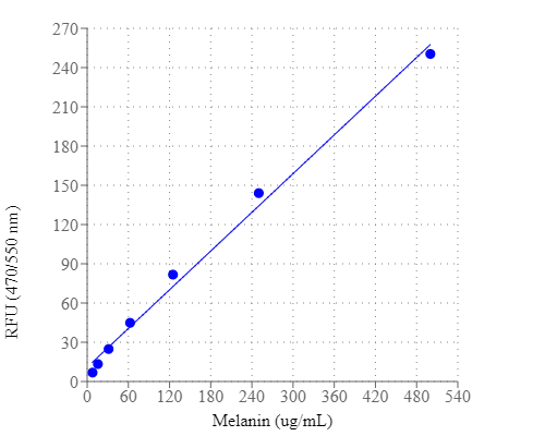
Amplite® Fluorimetric Melanin Assay Kit
Melanins have very diverse roles and functions in various organisms. Since melanins are an important biomarker, the accurate and sensitive determination of melanins has become a critical task for biomedical research and diagnostic applications. To address this unmet need, we have developed a robust fluorescence-based melanin assay. Amplite® Fluorimetric Melanin Assay Kit uses a substrate that generates a fluorescent product upon reaction with melanins. Its fluorescence intensity is proportional to the amount of melanins in a sample. Amplite® Fluorimetric Melanin Assay Kit provides a simple and effective method to measure melanin content in cells and other samples. The plate-based assay format is designed to use with a fluorescent microplate reader.
| Catalog | Size | Price | Quantity |
|---|---|---|---|
| 11310 | 100 Tests | Price |
Usage and storage
| Intended use | Research Use Only (RUO) |
Instrument settings
| Fluorescence microplate reader | |
| Excitation | 470 nm |
| Emission | 550 nm |
| Cutoff | 515 nm |
| Recommended plate | Solid black |
Contact us
| Telephone | |
| Fax | |
| sales@aatbio.com | |
| International | See distributors |
| Bulk request | Inquire |
| Custom size | Inquire |
| Technical Support | Contact us |
| Request quotation | Request |
| Purchase order | Send to sales@aatbio.com |
| Shipping | Standard overnight for United States, inquire for international |
Page updated on July 16, 2026