logo
AAT Bioquest

Amplite® Fluorimetric Melanin Assay Kit

Melanins have very diverse roles and functions in various organisms. Since melanins are an important biomarker, the accurate and sensitive determination of melanins has become a critical task for biomedical research and diagnostic applications. To address this unmet need, we have developed a robust fluorescence-based melanin assay. Amplite® Fluorimetric Melanin Assay Kit uses a substrate that generates a fluorescent product upon reaction with melanins. Its fluorescence intensity is proportional to the amount of melanins in a sample. Amplite® Fluorimetric Melanin Assay Kit provides a simple and effective method to measure melanin content in cells and other samples. The plate-based assay format is designed to use with a fluorescent microplate reader.
Example protocol

AT A GLANCE

Protocol Summary
  1. Prepare and add standards and samples (50 µL)
  2. Add Signal Enhancer to the standards and wells (50 µL)
  3. Incubate the plate at room temperature for 30 to 60 minutes
  4. Monitor the fluorescence intensity at Ex/Em= 470/550 nm 
Important      Bring all the kit components at room temperature before starting the experiment.

PREPARATION OF STOCK SOLUTIONS

Unless otherwise noted, all unused stock solutions should be divided into single-use aliquots and stored at -20 °C after preparation. Avoid repeated freeze-thaw cycles.

Melanin stock solution
Add 120 µL DMSO (Component D) into Melanin Standard (Component A) and mix well to generate a 5 mg/mL stock solution. Keep the mixture at room temperature for 10 minutes. Now the standard is ready to be used.
Note      If you observe undissolved matter at the bottom, centrifuge the tube at 1000 rpm for 5 mins and take the supernatant and use that as a Melanin Standard solution.
Note      Store the unused Melanin stock solution at -20 °C in single use aliquots.

PREPARATION OF STANDARD SOLUTION

For convenience, use the Serial Dilution Planner:
https://www.aatbio.com/tools/serial-dilution/11310


Melanin standard
Use Melanin stock solution and Assay Buffer to generate 500 µg/mL concentration of Melanin standard solution (M1). Then perform 1:2 serial dilutions to get remaining serially diluted Melanin standards (M2-M7). Note: The final in well concentration of the standards will be 1/2X.

SAMPLE EXPERIMENTAL PROTOCOL

Table 1.Layout of Melanin standards and test samples in a solid black 96- wells microplate. Melanin standards (M1-M7= 500 to 7.81 µg/mL), TS= Test Samples, BL= Blank Samples
BLBLTSTS
M1M1......
M2M2......
M3M3  
M4M4  
M5M5  
M6M6  
M7M7  
  1. Prepare the standards and test samples as per recommendations in assay buffer and add 50 µL of each in a microplate.
  2. Add 50 µL Signal Enhancer (Component C) to all the wells.
  3. Incubate the reaction at room temperature for 30 to 60 minutes.
  4. Monitor the fluorescence intensity with fluorescence plate reader at Ex/Em= 470/550 nm with cutoff= 515 nm. 

Citations

View all 10 citations: Citation Explorer
Nasal Cavity Administration of Melanin-Concentrating Hormone Improves Memory Impairment in Memory-Impaired and Alzheimer's Disease Mouse Models
Authors: Oh, S. T., Liu, Q. F., Jeong, H. J., Lee, S., Samidurai, M., Jo, J., Pak, S. C., Park, H. J., Kim, J., Jeon, S.
Journal: Mol Neurobiol (2019): ersion="1.0" encoding="UTF-8" ?>11310.enlEndN
The Melanin-concentrating Hormone System in Human, Rodent and Avian Brain
Authors: Bencze, J., Pocsai, K., Murnyak, B., Gergely, P. A., Juhasz, B., Szilvassy, Z., Hortobagyi, T.
Journal: Open Med (Wars) (2018): 264-269
Novel Neuroprotective Effects of Melanin-Concentrating Hormone in Parkinson's Disease
Authors: Park, J. Y., Kim, S. N., Yoo, J., Jang, J., Lee, A., Oh, J. Y., Kim, H., Oh, S. T., Park, S. U., Kim, J., Park, H. J., Jeon, S.
Journal: Mol Neurobiol (2017): 7706-7721
Melanin or a Melanin-Like Substance Interacts with the N-Terminal Portion of Prion Protein and Inhibits Abnormal Prion Protein Formation in Prion-Infected Cells
Authors: Hamanaka, T., Nishizawa, K., Sakasegawa, Y., Oguma, A., Teruya, K., Kurahashi, H., Hara, H., Sakaguchi, S., Doh-Ura, K.
Journal: J Virol (2017): se name="11310.enl" path="C:\Users\aatbi\Drop
Paraquat-Melanin Redox-Cycling: Evidence from Electrochemical Reverse Engineering
Authors: Kim, E., Leverage, W. T., Liu, Y., Panzella, L., Alfieri, M. L., Napolitano, A., Bentley, W. E., Payne, G. F.
Journal: ACS Chem Neurosci (2016): 1057-67
Page updated on December 13, 2024

Ordering information

Price
Unit size
Catalog Number11310
Quantity
Add to cart

Additional ordering information

Telephone1-800-990-8053
Fax1-800-609-2943
Emailsales@aatbio.com
InternationalSee distributors
Bulk requestInquire
Custom sizeInquire
Technical SupportContact us
Purchase orderSend to sales@aatbio.com
ShippingStandard overnight for United States, inquire for international
Request quotation

Storage, safety and handling

Intended useResearch Use Only (RUO)

Platform

Fluorescence microplate reader

Excitation470 nm
Emission550 nm
Cutoff515 nm
Recommended plateSolid black

Components

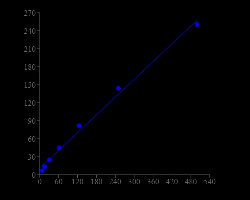
Melanin dose response was measured with Amplite® Fluorimetric Melanin Assay Kit in a 96-well black plate using a Gemini microplate reader (Molecular Devices). Equal volume of melanin standards and Signal Enhancer were added and incubated for 45 minutes at room temperature.  The signal was acquired at Ex/Em = 470/550 nm (cut off at 515 nm).