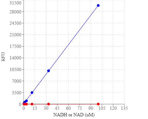
Amplite® Fluorimetric NADH Assay Kit *Red Fluorescence*
Price | |
Catalog Number | |
Unit Size | |
Quantity |
Telephone | 1-800-990-8053 |
Fax | 1-800-609-2943 |
sales@aatbio.com | |
International | See distributors |
Bulk request | Inquire |
Custom size | Inquire |
Shipping | Standard overnight for United States, inquire for international |
H-phrase | H303, H313, H333 |
Hazard symbol | XN |
Intended use | Research Use Only (RUO) |
R-phrase | R20, R21, R22 |
UNSPSC | 12352200 |
Overview | ![]() ![]() |
Platform
Fluorescence microplate reader
Excitation | 540 nm |
Emission | 590 nm |
Cutoff | 570 nm |
Recommended plate | Solid black |
Components
Example protocol
AT A GLANCE
Protocol summary
- Prepare NADH working solution (50 µL)
- Add NADH standards or test samples (50 µL)
- Incubate at room temperature for 15 minutes - 2 hours
- Monitor fluorescence intensity at Ex/Em = 540/590 nm
Important notes
Thaw one of each kit component at room temperature before starting the experiment.
PREPARATION OF STOCK SOLUTION
1. NADH standard solution (1 mM):
Add 200 µL of PBS buffer into the vial of NADH standard (Component C) to make 1 mM (1 nmol/µL) NADH standard solution.
PREPARATION OF STANDARD SOLUTION
For convenience, use the Serial Dilution Planner: https://www.aatbio.com/tools/serial-dilution/15261
Take the NADH standard solution and use PBS buffer to generate 100 µM NADH standard solution (NS7). Then perform 1:3 serial dilutions to get remaining serial dilutions of NADH standard (NS6 - NS1). Note: Diluted NADH standard solution is unstable, and should be used within 4 hours.
PREPARATION OF WORKING SOLUTION
Add 10 mL of Amplite™ NADH Assay Buffer (Component B) to the bottle of NADH Recycling Enzyme Mixture (Component A) and mix well. Note: This NADH working solution is enough for two 96-well or four 384-well plates. The working solution is not stable, use it promptly and avoid direct exposure to light.
SAMPLE EXPERIMENTAL PROTOCOL
Table 1. Layout of NADH standards and test samples in a solid black 96-well microplate. NS = NADH standard (NS1 - NS7, 0.1 to 100 µM); BL = blank control; TS = test sample.
BL | BL | TS | TS |
NS1 | NS1 | ... | ... |
NS2 | NS2 | ... | ... |
NS3 | NS3 | ||
NS4 | NS4 | ||
NS5 | NS5 | ||
NS6 | NS6 | ||
NS7 | NS7 |
Table 2. Reagent composition for each well.
Well | Volume | Reagent |
NS1 - NS7 | 50 µL | Serial Dilution (0.1 to 100 µM) |
BL | 50 µL | PBS |
TS | 50 µL | Test Sample |
- Prepare NADH standards (NS), blank controls (BL), and test samples (TS) according to the layout provided in Table 1 and Table 2. For a 384-well plate, use 25 µL of reagent per well instead of 50 µL.
- Add 50 µL of NADH working solution into each well of NADH standard, blank control, and test samples to make the total NADH assay volume of 100 µL/well. For a 384-well plate, add 25 µL of NADH working solution into each well instead, for a total volume of 50 µL/well.
- Incubate the reaction at room temperature for 15 minutes to 2 hours, protected from light.
- Monitor the fluorescence increase with a fluorescence plate reader at Excitation = 530 - 570 nm, Emission = 590 - 600 nm (optimal at Ex/Em = 540/590 nm, cutoff=570 nm). Note: The contents of the plate can also be transferred to a white clear bottom plate and read by an absorbance microplate reader at the wavelength of 576 ± 5 nm. However, the absorption detection will have a lower sensitivity compared to that of the fluorescence reading. For cell based NADH measurements, ReadiUse™ mammalian cell lysis buffer *5X* (Cat No. 20012) is recommended to use for lysing the cells.
Images
Citations
Authors: Allie, Robert
Journal: (2022)
Authors: Lu, Xinyao and Ren, Shunli and Lu, Jingzheng and Zong, Hong and Song, Jian and Zhuge, Bin
Journal: Journal of applied microbiology (2018)
Authors: Luo, Gang and Huang, Bingqing and Qiu, Xiang and Xiao, Lin and Wang, Ning and Gao, Qin and Yang, Wei and Hao, Liping
Journal: Molecular Nutrition & Food Research (2017)
Authors: Ling, Min and Huang, Peixin and Islam, Shamima and Heruth, Daniel P and Li, Xuanan and Zhang, Li Qin and Li, Ding-You and Hu, Zhaohui and Ye, Shui Qing
Journal: Cell & Bioscience (2017): 27
Authors: Ren, T and Zhang, H and Wang, J and Zhu, J and Jin, M and Wu, Y and Guo, X and Ji, L and Huang, Q and Yang, H and others, undefined
Journal: Oncogene (2017)
Authors: Zhang, Li Q and Van Ha, undefined and el, Leon and Xiong, Min and Huang, Peixin and Heruth, Daniel P and Bi, Charlie and Gaedigk, Roger and Jiang, Xun and Li, Ding-You and Wyckoff, Gerald and others, undefined
Journal: Cell Death & Disease (2017): e2705
Authors: Yang, Fei and Heit, Caitlin and Inglis, Debra L
Journal: Fermentation (2017): 61
Authors: Li, Miao and Liu, Xin and He, Yongpeng and Zheng, Qingyin and Wang, Min and Wu, Yu and Zhang, Yuanpeng and Wang, Chaoyun
Journal: European Journal of Pharmacology (2017): 124--133
Authors: Saihara, Kazuhiro and Kamikubo, Ryosuke and Ikemoto, Kazuto and Uchida, Koji and Akagawa, Mitsugu
Journal: Biochemistry (2017)
Authors: Wang, Chen and Cai, Heng and Chen, Zhongjun and Zhou, Zhihui
Journal: Biotechnology letters (2016): 1791--1797
Application notes
Acetylcholinesterase Inhibitory Activity of Pigment Echinochrome A
Ameliorative Effect of Novel Vitamin Formula with Herbal Extracts on Scopolamine-Induced Alzheimer's Disease
An Increase in Plasma Homovanillic Acid with Cocoa Extract Consumption Is Associated with the Alleviation of Depressive Symptoms in Overweight or Obese Adults
Attenuation of lysyl oxidase and collagen gene expression in keratoconus patient corneal epithelium corresponds to disease severity