logo
AAT Bioquest

Amplite® Fluorimetric NADP/NADPH Ratio Assay Kit *Red Fluorescence*

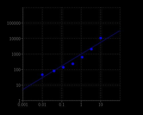
NADPH dose response was measured with Amplite® Fluorimetric NADP/NADPH Ratio Assay Kit in a 96-well solid black plate using a Gemini microplate reader (Molecular Devices).
NADPH/NADP+ and ATP contents of <em>Syn</em>, <em>Syn</em>/PFP, <em>R. palustris</em>, and <em>R. palustris</em>/PFP. NADP+/NADPH ratio found using Amplite fluorimetric assay kit 15264. Source: <b>Solar-powered multi-organism symbiont mimic system for beyond natural synthesis of polypeptides from CO2 and N2</b> by Wen Yu et al., <em>Science Advances</em> Vol. 9(11): eadf6772. March 2023.
Ordering information
Price
Catalog Number
Unit Size
Quantity
Add to cart
Additional ordering information
Telephone1-800-990-8053
Fax1-800-609-2943
Emailsales@aatbio.com
InternationalSee distributors
Bulk requestInquire
Custom sizeInquire
ShippingStandard overnight for United States, inquire for international
Request quotation
Storage, safety and handling
H-phraseH303, H313, H333
Hazard symbolXN
Intended useResearch Use Only (RUO)
R-phraseR20, R21, R22
UNSPSC12352200

OverviewpdfSDSpdfProtocol


Nicotinamide adenine dinucleotide (NAD+) and nicotinamide adenine dinucleotide phosphate (NADP+) are two important cofactors found in cells. NADH is the reduced form of NAD+, and NAD+ is the oxidized form of NADH. It forms NADP with the addition of a phosphate group to the 2' position of the adenyl nucleotide through an ester linkage. NADP is used in anabolic biological reactions, such as fatty acid and nucleic acid synthesis, which require NADPH as a reducing agent. The traditional NAD/NADH and NADP/NADPH assays are done by monitoring of NADH or NADPH absorption at 340 nm. This method suffers low sensitivity and high interference since the assay is done in the UV range that requires expensive quartz microplate. Our Amplite® NADP/NADPH Ratio Assay Kit provides a convenient method for sensitive detection of NADP, NADPH and their ratio. The enzymes in the system specifically recognize NAD/NADH in an enzyme cycling reaction. There is no need to purify NADP/NADPH from sample mix. The enzyme cycling reaction significantly increases detection sensitivity. In addition, this assay has very low background since it is run in the red visible range that significantly reduces the interference from biological samples. The assay has demonstrated high sensitivity and low interference. This Amplite® Fluorimetric NADP/NADPH Assay Kit can be performed in a convenient 96-well or 384-well microtiter-plate format and easily adapted to automation with no separation steps required.
Platform


Absorbance microplate reader

Absorbance576 ± 5 nm
Recommended plateClear bottom

Fluorescence microplate reader

Excitation540 nm
Emission590 nm
Cutoff570 nm
Recommended plateSolid black

Components


Example protocol


AT A GLANCE

Protocol Summary
  1. Prepare 25 µL NADPH standards and/or test samples
  2. Add 25 µL of NADPH or NADP Extraction Solution
  3. Incubate at 37oC for 15 minutes
  4. Add 25 µL of NADP or NADPH Extraction Solution
  5. Add 75 µL NADP/NADPH working solution
  6. Incubate at 37 °C for 15 minutes – 2 hours

  7. Monitor fluorescence intensity at Ex/Em = 540/590 nm (Cutoff = 570 nm)
Important Note

It is highly recommended to incubate the cells with Lysis Buffer (Component G) at 37oC and use the supernatant for the experiment.

Thaw one of each kit component at room temperature before starting the experiment.

CELL PREPARATION

For guidelines on cell sample preparation, please visit https://www.aatbio.com/resources/guides/cell-sample-preparation.html

PREPARATION OF STOCK SOLUTIONS

Unless otherwise noted, all unused stock solutions should be divided into single-use aliquots and stored at -20 °C after preparation. Avoid repeated freeze-thaw cycles

NADPH standard solution (1 mM)

Add 200 µL of PBS buffer into the vial of NADPH standard (Component C) to make 1 mM (1 nmol/µL) NADPH standard solution.

PREPARATION OF STANDARD SOLUTIONS

For convenience, use the Serial Dilution Planner:
https://www.aatbio.com/tools/serial-dilution/15264

NADPH standard
Add 10 µL of 1 mM NADPH standard solution to 990 µL PBS buffer (pH 7.4) to generate 10 µM (10 pmols/µL) NADPH standard solution (NS7). Take 10 µM NADPH standard solution and perform 1:3 serial dilutions in PBS to get serial dilutions of NADPH standard (NS6 - NS1). Note: Diluted NADPH standard solution is unstable, and should be used within 4 hours.

PREPARATION OF WORKING SOLUTION

Add 10 mL of NADPH Sensor Buffer (Component B) into the bottle of NADP/NADPH Recycling Enzyme Mixture (Component A) and mix well to make NADP/NADPH working solution.
Note        This NADP/NADPH working solution is enough for 125 assays in 96-well plate.

SAMPLE EXPERIMENTAL PROTOCOL

Table 1. Layout of NADPH standards and test samples in a solid black 96-well microplate. NS= NADPH Standards (NS1 - NS7, 0.01 to 10 µM); BL=Blank Control; TS=Test Samples; TS (NADPH) = Test Samples treated with NADPH Extraction Solution, then neutralized by NADP Extraction Solution; TS (NADP) = Test Samples treated with NADP Extraction Solution, then neutralized by NADPH Extraction Solution.

BLBLTSTSTS (NADPH)TS (NADPH)TS (NADP)TS (NADP)
NS1NS1..................
NS2NS2..................
NS3NS3
NS4NS4
NS5NS5
NS6NS6
NS7NS7

Table 2. Reagent composition for each well. Note: High concentration of NADPH (e.g., >100 µM, final concentration) may cause reduced fluorescence signal due to the over oxidation of NADPH sensor (to a non-fluorescent product).

NADPH StandardBlank Control

Test Sample (NADP+NADPH)

Test Sample (NADPH Extract)Test Sample (NADP Extract)
Serial Dilutions*: 25 μLPBS: 25 μLTest Sample: 25 μLTest Sample: 25 μLTest Sample: 25 μL
Component F: 25 μLComponent F: 25 μLComponent F: 25 μLComponent D: 25 μLComponent E: 25 μL
Incubate at 37 °C for 10 to 15 minutes
Component F: 25 μLComponent F: 25 μLComponent F: 25 μLComponent E: 25 μLComponent D: 25 μL
Total: 75 μLTotal: 75 μLTotal: 75 μLTotal: 75 μLTotal: 75 μL
  1. Prepare NADPH standards (NS), blank controls (BL), and test samples (TS) according to the layout provided in Tables 1 and 2.

    Note        Prepare cells or tissue samples as desired. NADP/NADPH Lysis Buffer (Component G) can be used for lysing the cells for convenience. One can simply use total NADP and NADPH minus the NADP to calculate the amount of NADPH.

    Note        It is highly recommended to incubate the cells at 37 °C and use the supernatant for the experiment.
  2. For NADP Extraction (NADP): Add 25 µL of NADP Extraction Solution (Component E) into the wells of NADP/NADPH containing test samples. Incubate at 37 °C for 10 to 15 minutes, then add 25 µL of NADPH Extraction Solution (Component D) to neutralize the NADP extracts as described in Tables 1 & 2.For Total NADP and NADPH: Add 25 µL of NADP/NADPH Control Solution (Component F) into the wells of NADPH standards and NADP/NADPH containing test samples. Incubate at 37 °C for 10 to 15 minutes, and then add 25 µL of Control Solution (Component F) as described in Tables 1 & 2.For NADPH Extraction (NADPH): Add 25 µL of NADPH Extraction Solution (Component D) into the wells of NADP/NADPH containing test samples. Incubate at 37 °C for 10 to 15 minutes, then add 25 µL of NADP Extraction Solution (Component E) to neutralize the NADPH extracts as described in Tables 1 & 2.
  3. Add 75 µL of NADPH working solution into each well of NADPH standard, blank control, and test samples to make the total NADPH assay volume of 150 µL/well.
  4. Incubate the reaction at 37 °C for 15 minutes to 2 hours (We tested 60 minutes in the figure shown), protected from light.

    Note        In some cases, incubation time can be increased to more than 2 hours.
  5. Monitor the fluorescence increase with a fluorescence plate reader at Ex/Em = 540/590 nm. Note: The contents of the plate can also be transferred to a white clear bottom plate and read by an absorbance microplate reader at the wavelength of 576 ± 5 nm. The absorption detection has lower sensitivity compared to fluorescence reading.

Images


Citations


View all 81 citations: Citation Explorer
Solar-powered multi-organism symbiont mimic system for beyond natural synthesis of polypeptides from CO2 and N2
Authors: Yu, Wen and Zeng, Yue and Wang, Zenghao and Xia, Shengpeng and Yang, Zhiwen and Chen, Weijian and Huang, Yiming and Lv, Fengting and Bai, Haotian and Wang, Shu
Journal: Science Advances (2023): eadf6772
The combined effect of nitrogen deprivation and overexpression of malic enzyme gene on growth and lipid accumulation in Phaeodactylum tricornutum
Authors: Zhang, Ruihao and Zhu, Baohua and Tu, Changchao and Li, Yun and Zhao, Yan and Pan, Kehou
Journal: Journal of Applied Phycology (2021): 1--9
Artificial regulation of state transition for augmenting plant photosynthesis using synthetic light-harvesting polymer materials
Authors: Zhou, Xin and Zeng, Yue and Tang, Yongyan and Huang, Yiming and Lv, Fengting and Liu, Libing and Wang, Shu
Journal: Science advances (2020): eabc5237
Calcitonin gene-related peptide inhibits angiotensin II-induced NADPH oxidase-dependent ROS via the Src/STAT3 signalling pathway
Authors: Luo, Hong-min and Wu, Xia and Xian, Xian and Wang, Lu-yao and Zhu, Liang-yu and Sun, Hong-yu and Yang, Lei and Liu, Wen-xuan
Journal: Journal of Cellular and Molecular Medicine (2020)
Growth suppression by altered (p) ppGpp levels results from non-optimal resource allocation in Escherichia coli
Authors: Zhu, Manlu and Dai, Xiongfeng
Journal: Nucleic acids research (2019)
6-Phosphogluconate Dehydrogenase Links Cytosolic Carbohydrate Metabolism to Protein Secretion via Modulation of Glutathione Levels
Authors: Li, Haoxin and Ericsson, Maria and Rabasha, Bokang and Budnik, Bogdan and Chan, Sze Ham and Freinkman, Elizaveta and Lewis, Caroline A and Doench, John G and Wagner, Bridget K and Garraway, Levi A and others, undefined
Journal: Cell chemical biology (2019)
A metabolomic study of the effect of Candida albicans glutamate dehydrogenase deletion on growth and morphogenesis
Authors: Han, Ting-Li and Cannon, Richard D and Gallo, S and ra M , undefined and Villas-Boas, Silas G
Journal: NPJ biofilms and microbiomes (2019): 13
The role of malic enzyme on promoting total lipid and fatty acid production in Phaeodactylum tricornutum
Authors: Zhu, Bao-Hua and Zhang, Rui-Hao and Lv, Na-Na and Yang, Guan-Pin and Wang, Yi-Sheng and Pan, Ke-Hou
Journal: Frontiers in plant science (2018): 826
A cytosolic triosephosphate isomerase is a key component in XA3/XA26-mediated resistance
Authors: Liu, Yanyan and Cao, Yinglong and Zhang, Qinglu and Li, Xianghua and Wang, Shiping
Journal: Plant physiology (2018): 923--935