logo
AAT Bioquest

Buccutite™ Poly-HRP Antibody Conjugation Kit

Poly-HRP-antibody conjugates is considered to have the highest sensitivity in immunoassays where the sample is limited, or a target molecule is present at extremely low level. Buccutite™ Poly-HRP Antibody Conjugation Kit is designed for rapid preparation of poly-HRP conjugated antibodies. The kit provides the poly-HRP that is pre-activated with our proprietary linker Buccutite™ FOL. A targeted antibody is activated with Buccutite™ MTA (provided in the kit) to give MTA-activated antibody. The simple mix of MTA-activated antibody with FOL-activated poly-HRP to give the desired poly-HRP-labeled antibody conjugate. The reaction occurs under extremely mild conditions without a catalyst required. The poly-HRP antibody conjugate prepared with Buccutite™ Poly-HRP Antibody Conjugation Kit is compatible with chromogenic, fluorogenic and chemiluminescent HRP substrates used in ELISA, western blotting, immunohistochemistry (IHC) assays. It can also be used with TSA or our Styramide™ fluorescent HRP substrates for ultrasensitive detection of low abundant biological targets.

Example protocol

AT A GLANCE

Protocol summary
  1. Add 5 µL of Reaction Buffer (Component C) into antibody (50 µL)

  2. Add the antibody solution into the Buccutite™ MTA vial (Component B)
  3. Incubate at room temperature for 30 minutes
  4. Mix with 200 µL of Buccutite™ FOL-Activated Poly-HRP (Component A)
  5. Incubate at room temperature for 60 minutes
Important

Upon receipt, store the kit at 4 °C. When stored properly, the kit should be stable for six months. Alternatively, Component B can be stored at -20 °C. Warm up all the Components to room temperature and centrifuge the vials briefly before opening. Immediately prepare the required solutions before starting your conjugation. The following SOP is an example for labeling 50 µg goat anti-mouse IgG antibody.

PREPARATION OF WORKING SOLUTION

Antibody working solution

For labeling 50 µg antibody (assuming the target antibody concentration is 1 mg/mL), mix 5 µL (5% of the total reaction volume) of Reaction Buffer (Component C) with 50 µL of the target antibody solution.
Note        If you have a different concentration, adjust the antibody volume accordingly to make ~50 µg antibody/50 µL available for your labeling reaction.
Note        The antibody should be dissolved in 1X phosphate buffered saline (PBS), pH 7.2-7.4. If the antibody is dissolved in glycine buffer, it must be dialyzed against 1X PBS, pH 7.2-7.4, or use 10KD Filter (Cat. # 60502 from AAT Bioquest) to remove free amines or ammonium salts (such as ammonium sulfate and ammonium acetate).
Note        Impure antibodies or antibodies stabilized with bovine serum albumin (BSA) or gelatin will not be labeled well.
Note        The antibody –Buccutite™ MTA reaction efficiency is significantly reduced if the antibody concentration is less than 1 mg/mL. For optimal labeling efficiency, the final antibody concentration range of 1-10 mg/mL is recommended.

SAMPLE EXPERIMENTAL PROTOCOL

Run Antibody-Buccutite™ MTA reaction
  1. Add the antibody working solution directly into the vial of Buccutite ™ MTA (Component B), and mix them well by repeatedly pipetting for a few times or vortex the vial for a few seconds.
  2. Incubate the antibody-Buccutite ™ MTA reaction mixture at room temperature for 30 minutes.

    Note        The antibody-Buccutite™ MTA reaction mixture can be rotated or shaken for longer time if desired.
Make HRP-antibody conjugation
  1. Add antibody-Buccutite™ MTA reaction mixture to the vial of Buccutite™ FOL-Activated Poly-HRP (Component A), mix well and incubate the mixture at room temperature for 60 minutes.

    Note        The antibody poly-HRP reaction mixture can be incubated for longer time if desired.
  2. The antibody-poly-HRP conjugate is now ready to use.

    Note        For immediate use, the antibody-poly-HRP conjugate needs to be diluted with the buffer of your choice.
  3. The concentration of the conjugate can be calculated as follows: Antibody Concentration (µg/µL) = 50 µg (total amount of antibody)/(50 µL + 5 µL+ 200 µL) = 0.196 µg/µL
Storage of Antibody-Poly-HRP Conjugate

The antibody conjugate-polyHRP conjugate should be stored in the presence of a carrier antibody (e.g., 0.1% bovine serum albumin) at 4 °C and kept from light for two months.

References

View all 11 references: Citation Explorer
Detecting cancer metastasis and accompanying protein biomarkers at single cell levels using a 3D-printed microfluidic immunoarray.
Authors: Sharafeldin, Mohamed and Chen, Tianqi and Ozkaya, Gulsum Ucak and Choudhary, Dharamainder and Molinolo, Alfredo A and Gutkind, J Silvio and Rusling, James F
Journal: Biosensors & bioelectronics (2021): 112681
Prostate Cancer Diagnosis in the Clinic Using an 8-Protein Biomarker Panel.
Authors: Jones, Abby L and Dhanapala, Lasangi and Baldo, Thaísa A and Sharafeldin, Mohamed and Krause, Colleen E and Shen, Min and Moghaddam, Shirin and Faria, Ronaldo C and Dey, Dipak K and Watson, R William and Andrawis, Ramez and Lee, Norman H and Rusling, James F
Journal: Analytical chemistry (2020)
Comparison of different methodologies and cryostat versus paraffin sections for chromogenic immunohistochemistry.
Authors: Hira, Vashendriya V V and de Jong, Annique Loncq and Ferro, Klea and Khurshed, Mohammed and Molenaar, Remco J and Van Noorden, Cornelis J F
Journal: Acta histochemica (2019): 125-134
The complementary role of insulin-like growth factor II mRNA-binding protein 3 (IMP3) in diagnosis of Hodgkin's lymphoma.
Authors: Masoud, Rokia and Ibrahiem, Afaf and Tantawy, Dina and Eldosoky, Ibrahim
Journal: Annals of diagnostic pathology (2019): 64-68
Using magnetic beads and signal amplifiers to produce short and simple immunoassays: Application to MMP-9 detection in plasma samples.
Authors: Ben Ismail, Manel and de la Serna, Erica and Ruiz-Vega, Gisela and García-Berrocoso, Teresa and Montaner, Joan and Zourob, Mohammed and Othmane, Ali and Baldrich, Eva
Journal: Analytica chimica acta (2018): 144-154
Page updated on December 11, 2024

Ordering information

Price
Unit size
1x50 ug Labeling
2x50 ug Labelings
Catalog Number
55185519
Quantity
Add to cart

Additional ordering information

Telephone1-800-990-8053
Fax1-800-609-2943
Emailsales@aatbio.com
InternationalSee distributors
Bulk requestInquire
Custom sizeInquire
Technical SupportContact us
Purchase orderSend to sales@aatbio.com
ShippingStandard overnight for United States, inquire for international
Request quotation

Storage, safety and handling

H-phraseH303, H313, H333
Hazard symbolXN
Intended useResearch Use Only (RUO)
R-phraseR20, R21, R22
UNSPSC12171501

Components

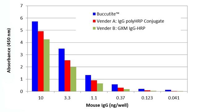
Direct ELISA curves were generated using the polyHRP- goat-anti-mouse IgG conjugate prepared with Buccutite&trade; Poly-HRP Antibody Conjugation Kit (Cat No. 5518, Cat No. 5519). A 3-fold&nbsp; serial diluted mouse IgG was coated onto a 96-well plate, and 100 &micro;L GAM IgG-polyHRP conjugate (100 ng/ml) was tested using the standard ELISA method. TMB substrate solution (Cat No. 11003) was used to detect the immobilized mouse IgG with 5 min incubation and read at 450 nm.<br><br />Blue: Buccutite&trade; Kit 5518 or 5519<br />Red: GXM IgG PolyHRP (Vendor A)<br />Green: GXM IgG-HRP (Vendor B)
ELISA curves were generated using the polyHRP- goat-anti-mouse IgG conjugate prepared with Buccutite™ Poly-HRP Antibody Conjugation Kit ( Cat#5518 or Cat#5519). 3-fold  serial diluted mouse IgG was coated on a 96-well plate, and  100uL GAM IgG-polyHRP conjugate (100ng/ml) was tested using the standard ELISA method. TMB substrate solution (Cat#11003) was used to detect the immobilized mouse IgG with 5 min incubation and read at 450 nm.