logo
AAT Bioquest

Cell Meter™ Caspase 8 Activity Apoptosis Assay Kit *Green Fluorescence*

Our Cell Meter™ assay kits are a set of tools for monitoring cellular functions. There are a variety of parameters that can be used to monitor cell apoptosis. This particular kit is designed to monitor cell apoptosis by measuring caspase 8 activity. Caspase 8 is a caspase protein, encoded by the CASP8 gene. Caspase 8 also plays an important role in neurodegenerative diseases such as Huntington disease. Caspase 8 is proven to have substrate selectivity for the peptide sequence Ile-Glu-Thr-Asp (IETD). This kit uses (Ac-IETD)2-R110 as a fluorogenic indicator for caspase 8 activity. Cleavage of rhodamine 110 (R110) peptides by caspase 8 generates strongly fluorescent R110 which is monitored at the emission between 520 nm and 530 nm with the excitation between 480 nm and 500 nm. This spectral feature makes the kit compatible with the FITC filter set. The kit provides all the essential components with an optimized assay protocol. The assay can be readily adapted for high throughput screenings. It can be used to either quantify the activated caspase 8 activities in apoptotic cells or screen the caspase 8 inhibitors.
Example protocol

AT A GLANCE

Protocol summary

  1. Prepare cells with test compounds (100 µL/well/96-well plate or 25 µL/well/384-well plate)
  2. Add equal volume of Caspase 8 Substrate working solution (100 µL/well/96-well plate or 25 µL/well/384-well plate)
  3. Incubate at room temperature for 30 min to 1 hour
  4. Monitor fluorescence increase at Ex/Em = 490/525 nm (Cutoff = 515 nm)

Important notes
Thaw one vial of each kit component at room temperature before starting the experiment.

PREPARATION OF WORKING SOLUTION

Add 50 µL of Caspase 8 Substrate (Component A) into 10 mL of Assay Buffer (Component B) and mix well to make Caspase 8 Substrate working solution. Protect from light. Note: Caspasae 8 Substrate working solution is not stable, use it promptly. Note: Aliquot and store unused Caspase 8 Substrate (Component A) and Assay Buffer (Component B) at -20 oC. Avoid repeated freeze/thaw cycles.

For guidelines on cell sample preparation, please visit
https://www.aatbio.com/resources/guides/cell-sample-preparation.html

SAMPLE EXPERIMENTAL PROTOCOL

  1. Treat cells by adding 10 µL/well of 10X test compounds (96-well plate) or 5 µL/well of 5X test compounds (384-well plate) into PBS or the desired buffer. For blank wells (medium without the cells), add the same amount of compound buffer.

  2. Incubate the cell plate in a 5% CO2, 37°C incubator for a desired period of time (4 - 6 hours for Jurkat cells treated with camptothecin or staurosporine) to induce apoptosis.

  3. Add 100 µL/well (96-well plate) or 25 µL/well (384-well plate) of Caspase 8 Substarte working solution.

  4. Incubate the plate at room temperature for 30 min to 1 hour, protected from light. Note: If desired, add 1 µL of the 1 mM Ac-IETD-CHO caspase 8 inhibitor to selected samples 10 minutes before adding Caspase 8 Substrate working solution at room temperature to confirm the inhibition of the caspase 8-like activities.

  5. Centrifuge cell plate (especially for the non-adherent cells) at 800 rpm for 2 minutes (brake off).

  6. Monitor the fluorescence increase with a fluorescence microplate reader at Ex/Em = 490/525 nm (Cutoff = 515 nm).

Spectrum

Citations

View all 19 citations: Citation Explorer
Contact-dependent signaling triggers tumor-like proliferation of CCM3 knockout endothelial cells in co-culture with wild-type cells
Authors: Rath, Matthias and Schwefel, Konrad and Malinverno, Matteo and Skowronek, Dariush and Leopoldi, Alexandra and Pilz, Robin A and Biedenweg, Doreen and Bekeschus, Sander and Penninger, Josef M and Dejana, Elisabetta and others,
Journal: Cellular and Molecular Life Sciences (2022): 1--20
Combination of Goniothalamin and Sol-Gel-Derived Bioactive Glass 45S5 Enhances Growth Inhibitory Activity via Apoptosis Induction and Cell Cycle Arrest in Breast Cancer Cells MCF-7
Authors: Bakar, Siti Aishah Abu and Ali, Abdul Manaf and Noor, Siti Noor Fazliah Mohd and Hamid, Shahrul Bariyah Sahul and Azhar, Nur Asna and Mohamad, Noor Muzamil and Ahmad, Nor Hazwani
Journal: BioMed Research International (2022)
RPL21 siRNA blocks proliferation in pancreatic cancer cells by inhibiting DNA replication and inducing G1 arrest and apoptosis
Authors: Li, Chaodong and Ge, Mei and Chen, Daijie and Sun, Tao and Jiang, Hua and Xie, Yueqing and Lu, Huili and Zhang, Baohong and Han, Lei and Chen, Junsheng and others,
Journal: Frontiers in oncology (2020): 1730
CD5L-induced activation of autophagy is associated with hepatoprotection in ischemic reperfusion injury via the CD36/ATG7 axis
Authors: Li, Junjian and Lin, Wei and Zhuang, Lei
Journal: Experimental and Therapeutic Medicine (2020): 2588--2596

References

View all 31 references: Citation Explorer
Discovery of substituted N'-(2-oxoindolin-3-ylidene)benzohydrazides as new apoptosis inducers using a cell- and caspase-based HTS assay
Authors: Sirisoma N, Pervin A, Drewe J, Tseng B, Cai SX.
Journal: Bioorg Med Chem Lett (2009): 2710
Discovery of 1-benzoyl-3-cyanopyrrolo[1,2-a]quinolines as a new series of apoptosis inducers using a cell- and caspase-based high-throughput screening assay. 2: Structure-activity relationships of the 4-, 5-, 6-, 7- and 8-positions
Authors: Kemnitzer W, Kuemmerle J, Jiang S, Sirisoma N, Kasibhatla S, Crogan-Grundy C, Tseng B, Drewe J, Cai SX.
Journal: Bioorg Med Chem Lett (2009): 3481
Selective identification of cooperatively binding fragments in a high-throughput ligation assay enables development of a picomolar caspase-3 inhibitor
Authors: Schmidt MF, El-Dahshan A, Keller S, Rademann J.
Journal: Angew Chem Int Ed Engl (2009): 6346
Discovery of 4-aryl-4H-chromenes as a new series of apoptosis inducers using a cell- and caspase-based high throughput screening assay. 4. Structure-activity relationships of N-alkyl substituted pyrrole fused at the 7,8-positions
Authors: Kemnitzer W, Drewe J, Jiang S, Zhang H, Crogan-Grundy C, Labreque D, Bubenick M, Attardo G, Denis R, Lamothe S, Gourdeau H, Tseng B, Kasibhatla S, Cai SX.
Journal: J Med Chem (2008): 417
Modified caspase-3 assay indicates correlation of caspase-3 activity with immunity of nonhuman primates to Yersinia pestis infection
Authors: Welkos S, Norris S, Adamovicz J.
Journal: Clin Vaccine Immunol (2008): 1134
Page updated on September 8, 2024

Ordering information

Price
Unit size
Catalog Number22798
Quantity
Add to cart

Additional ordering information

Telephone1-800-990-8053
Fax1-800-609-2943
Emailsales@aatbio.com
InternationalSee distributors
Bulk requestInquire
Custom sizeInquire
Technical SupportContact us
Purchase orderSend to sales@aatbio.com
ShippingStandard overnight for United States, inquire for international
Request quotation

Spectral properties

Extinction coefficient (cm -1 M -1)

80000

Excitation (nm)

500

Emission (nm)

522

Storage, safety and handling

H-phraseH303, H313, H333
Hazard symbolXN
Intended useResearch Use Only (RUO)
R-phraseR20, R21, R22
UNSPSC12352200

Platform

Fluorescence microplate reader

Excitation490 nm
Emission525 nm
Cutoff515 nm
Recommended plateBlack wall, clear bottom
Instrument specification(s)Top, Bottom read mode

Components

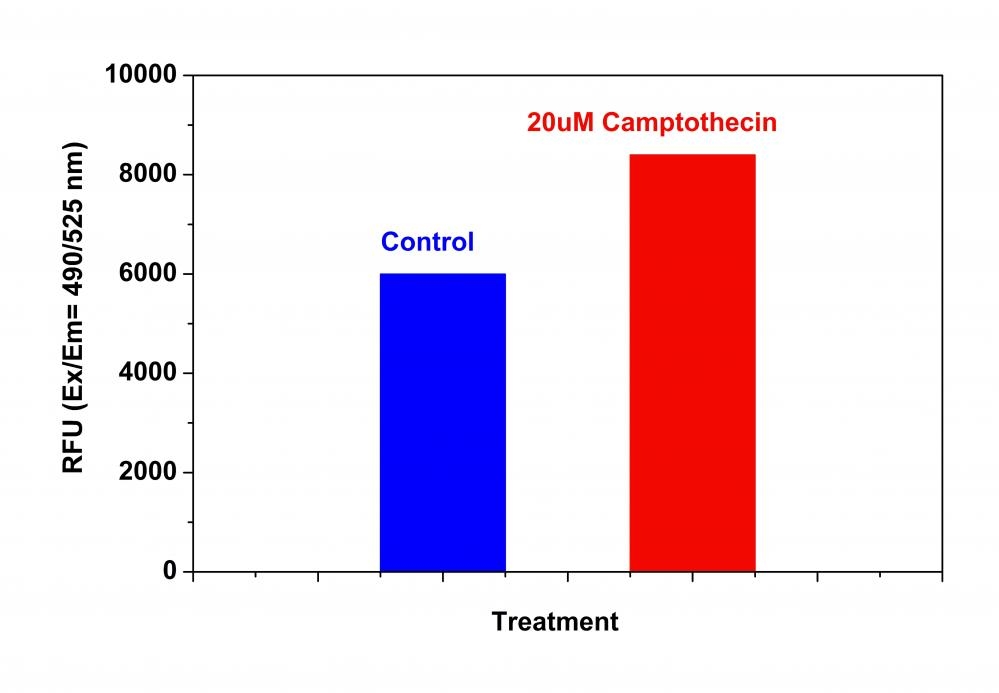
Detection of caspase 8 activities in Jurkat cells using Cell Meter™ Caspase 8 Activity Apoptosis Assay Kit. Jurkat cells were seeded on the same day at 200,000 cells/90 µL/well in a Costar black wall/clear bottom 96-well plate. The cells were treated with or without 20 µM of camptothecin for 4 hours. The caspase 8 Substrate working solution (100 µL/well) was added and incubated at room temperature for 1 hour. The fluorescence intensity was measured at Ex/Em = 490/525 nm (Cutoff = 515 nm) with a FlexStation™ microplate reader (Molecular Devices).