Cell Meter™ Cell Adhesion Assay Kit
Ordering information
Price | |
Catalog Number | |
Unit Size | |
Quantity |
Additional ordering information
Telephone | 1-800-990-8053 |
Fax | 1-800-609-2943 |
sales@aatbio.com | |
Quotation | Request |
International | See distributors |
Shipping | Standard overnight for United States, inquire for international |
Spectral properties
Excitation (nm) | 494 |
Emission (nm) | 514 |
Storage, safety and handling
H-phrase | H303, H313, H333 |
Hazard symbol | XN |
Intended use | Research Use Only (RUO) |
R-phrase | R20, R21, R22 |
UNSPSC | 12171501 |
Related products
Overview | ![]() ![]() |
Excitation (nm) 494 | Emission (nm) 514 |
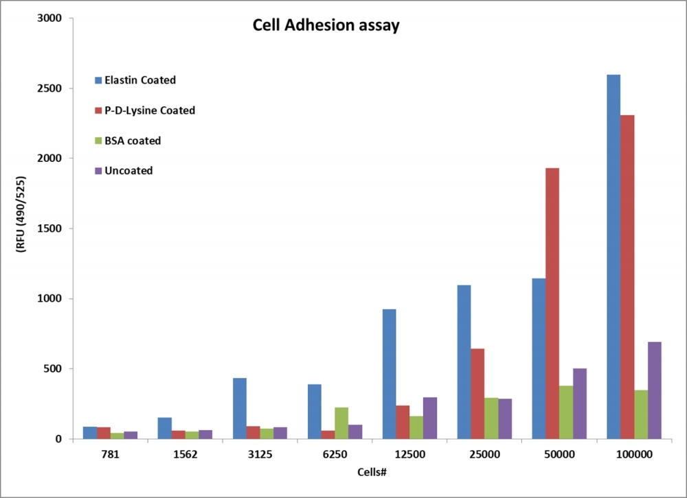
The Cell Meter™ Cell Adhesion Assay Kit is a fast and sensitive assay for measuring cell-cell or cell-surface adhesion for a variety of cell types. In this assay, cells are labeled with Calcein UltraGreen AM and allowed to adhere. After removal of nonadherent cells, The fluorescence of Calcein UltraGreen is used to calculate the number of adherent cells. The use of our outstanding fluorogenic dye, Calcein UltraGreen AM provides a fast and sensitive method for measuring cell adhesion with a variety of cell types. Calcein UltraGreen AM is nonfluorescent but, once loaded into cells, is cleaved by endogenous esterases to produce highly fluorescent Calcein UltraGreen, a brightly fluorescent, pH-independent, cytoplasmic cell marker with the minimal interference to cell adhesion process. The Cell Meter™ cell adhesion assay is designed for use with fluorescence microplate readers. The robust performance of Calcein UltraGreen AM and simple procedure of the kit avoids problems associated with assays that utilize radioisotopes, which generate hazardous waste, and with assays that rely on the use of covalently coupled cell-surface labels, which can potentially alter cell function.
Platform
Fluorescence microscope
Excitation | FITC filter set |
Emission | FITC filter set |
Recommended plate | Black wall/clear bottom |
Fluorescence microplate reader
Excitation | 490 nm |
Emission | 525 nm |
Cutoff | 515 nm |
Recommended plate | Black wall/Clear bottom |
Components
Example protocol
AT A GLANCE
Protocol summary
- Add cells on a plate coated with desired coating material
- Incubate cells at 37 °C to allow them to attach
- Remove the unattached cells
- Add Calcein Ultragreen AM working solution
- Incubate the cells at 37 °C for 20-30 minutes
- Remove supernatant and wash cells with HHBS or DPBS
- Measure the fluorescence intensity using fluorescence microplate reader with Ex/Em = 490/525 nm
Important
Thaw all the kit components at room temperature before starting the experiment.PREPARATION OF STOCK SOLUTIONS
Unless otherwise noted, all unused stock solutions should be divided into single-use aliquots and stored at -20 °C after preparation. Avoid repeated freeze-thaw cycles.
Note Store the unused Calcein Ultragreen AM stock solution at -20 °C in single use aliquots to avoid freeze thaw cycles.
Calcein Ultragreen AM stock solution
Add 50 µL of DMSO (Component C) into Calcein Ultragreen AM (Component A) and mix well.Note Store the unused Calcein Ultragreen AM stock solution at -20 °C in single use aliquots to avoid freeze thaw cycles.
PREPARATION OF WORKING SOLUTION
Calcein Ultragreen AM working solution
Add 50 µL of Calcein Ultragreen AM stock solution into 10 mL of Adhesion Assay Buffer and mix well.Note Calcein Ultragreen AM working solution should not be stored and should be used promptly.
Note 10 mL Calcein Ultragreen AM working solution is enough for 100 tests.
SAMPLE EXPERIMENTAL PROTOCOL
The following protocol can be used as a guideline and should be optimized according to the needs.
- Add 100 µL volumes of cells on a plate coated with desired coating material.
- Incubate plate at 37 °C for 2 to 3 hours.
Note For each cell line, optimal incubation time should be tested experimentally. - Remove the growth medium and unattached cells.
- Add 100 µL of Calcein Ultragreen AM working solution and incubate plate at 37 °C for 20-30 minutes.
Note For each cell line, optimal incubation time should be tested experimentally. - Remove the dye working solution and wash cells with 1X Hank’s salt solution and 20 mM Hepes buffer or DPBS once.
- Add 100 µL of Adhesion Assay Buffer to the wells.
- Monitor the fluorescence intensity using a fluorescence microplate reader at Ex/Em = 490/525 nm (Cutoff = 515 nm).
Product Family
Name | Excitation (nm) | Emission (nm) |
Cell Meter™ Cell Viability Assay Kit *Blue Fluorescence with 405 nm Excitation* | 406 | 445 |
Cell Meter™ Cell Viability Assay Kit *Green Fluorescence* | 494 | 514 |
Cell Meter™ Cell Proliferation Assay Kit | 503 | 527 |
Images

Figure 1. Cell adhesion measured with Cell Meter™ Cell Adhesion Assay Kit using a fluorescence microplate reader. Jurkat cells at different confluences or confluency levels were incubated in wells coated with different materials, and then stained with Calcein Ultragreen AM at 37°C for 30 mins. The signal was monitored at Ex/Em = 490/525 nm.

Figure 2. Effects of Hpgd on the adhesion and angiogenesis of hPAECs. (A) The microscope results showed that OE-Hpgd reduced the adhesion of hPAECs. (B) Statistical histogram of the adherent cells. (C) The microscope results showed that OE-Hpgd reduced the angiogenesis capability. (D) Statistical bar chart of the number of tubules. (**P<0.01). Source: Hpgd affects the progression of hypoxic pulmonary hypertension by regulating vascular remodeling by Meng He, Kelong Tao, Min Xiang & Jian Sun. BMC Pulmonary Medicine volume 23, Article number: 116. April 2023.
Citations
View all 1 citations: Citation Explorer
Hpgd affects the progression of hypoxic pulmonary hypertension by regulating vascular remodeling
Authors: He, Meng and Tao, Kelong and Xiang, Min and Sun, Jian
Journal: BMC Pulmonary Medicine (2023): 1--12
Authors: He, Meng and Tao, Kelong and Xiang, Min and Sun, Jian
Journal: BMC Pulmonary Medicine (2023): 1--12
References
View all 50 references: Citation Explorer
The Protein A-mediated binding of Staphylococcus to antibodies in flow cytometric assays and its reduction using FcR blocking reagent.
Authors: Cronin, Ultan P and Girardeaux, Laura and O'Meara, Elaine and Wilkinson, Martin
Journal: Applied and environmental microbiology (2020)
Authors: Cronin, Ultan P and Girardeaux, Laura and O'Meara, Elaine and Wilkinson, Martin
Journal: Applied and environmental microbiology (2020)
Effect of advanced glycation end product on paraoxonase 2 expression: Its impact on endoplasmic reticulum stress and inflammation in HUVECs.
Authors: Ravi, Ramya and Ragavachetty Nagaraj, Nareshkumar and Subramaniam Rajesh, Bharathidevi
Journal: Life sciences (2020): 117397
Authors: Ravi, Ramya and Ragavachetty Nagaraj, Nareshkumar and Subramaniam Rajesh, Bharathidevi
Journal: Life sciences (2020): 117397
Functionalized Graphene Nanoparticles Induce Human Mesenchymal Stem Cells to Express Distinct Extracellular Matrix Proteins Mediating Osteogenesis.
Authors: Newby, Steven D and Masi, Tom and Griffin, Christopher D and King, William J and Chipman, Anna and Stephenson, Stacy and Anderson, David E and Biris, Alexandru S and Bourdo, Shawn E and Dhar, Madhu
Journal: International journal of nanomedicine (2020): 2501-2513
Authors: Newby, Steven D and Masi, Tom and Griffin, Christopher D and King, William J and Chipman, Anna and Stephenson, Stacy and Anderson, David E and Biris, Alexandru S and Bourdo, Shawn E and Dhar, Madhu
Journal: International journal of nanomedicine (2020): 2501-2513
Apolipoprotein M and sphingosine-1-phosphate complex alleviates TNF-α-induced endothelial cell injury and inflammation through PI3K/AKT signaling pathway.
Authors: Liu, Yang and Tie, Li
Journal: BMC cardiovascular disorders (2019): 279
Authors: Liu, Yang and Tie, Li
Journal: BMC cardiovascular disorders (2019): 279
Angiotensin II Type I Receptor Blockade Is Associated with Decreased Cutaneous Scar Formation in a Rat Model.
Authors: Murphy, Amanda and LeVatte, Terry and Boudreau, Colton and Midgen, Craig and Gratzer, Paul and Marshall, Jean and Bezuhly, Michael
Journal: Plastic and reconstructive surgery (2019): 803e-813e
Authors: Murphy, Amanda and LeVatte, Terry and Boudreau, Colton and Midgen, Craig and Gratzer, Paul and Marshall, Jean and Bezuhly, Michael
Journal: Plastic and reconstructive surgery (2019): 803e-813e
Viable cryopreserved umbilical tissue (vCUT) reduces post-operative adhesions in a rabbit abdominal adhesion model.
Authors: Dhall, Sandeep and Coksaygan, Turhan and Hoffman, Tyler and Moorman, Matthew and Lerch, Anne and Kuang, Jin-Qiang and Sathyamoorthy, Malathi and Danilkovitch, Alla
Journal: Bioactive materials (2019): 97-106
Authors: Dhall, Sandeep and Coksaygan, Turhan and Hoffman, Tyler and Moorman, Matthew and Lerch, Anne and Kuang, Jin-Qiang and Sathyamoorthy, Malathi and Danilkovitch, Alla
Journal: Bioactive materials (2019): 97-106
Cardiomyocyte-myofibroblast contact dynamism is modulated by connexin-43.
Authors: Schultz, Francisca and Swiatlowska, Pamela and Alvarez-Laviada, Anita and Sanchez-Alonso, Jose L and Song, Qianqian and de Vries, Antoine A F and Pijnappels, Daniël A and Ongstad, Emily and Braga, Vania M M and Entcheva, Emilia and Gourdie, Robert G and Miragoli, Michele and Gorelik, Julia
Journal: FASEB journal : official publication of the Federation of American Societies for Experimental Biology (2019): 10453-10468
L-sepiapterin restores SLE serum-induced markers of endothelial function in endothelial cells.
Authors: Jones Buie, Joy N and Pleasant Jenkins, Dorea and Muise-Helmericks, Robin and Oates, Jim C
Journal: Lupus science & medicine (2019): e000294
Authors: Jones Buie, Joy N and Pleasant Jenkins, Dorea and Muise-Helmericks, Robin and Oates, Jim C
Journal: Lupus science & medicine (2019): e000294
Allogeneic platelet rich plasma serves as a scaffold for articular cartilage derived chondroprogenitors.
Authors: Vinod, Elizabeth and Vinod Francis, Deepak and Manickam Amirtham, Soosai and Sathishkumar, Solomon and Boopalan, P R J V C
Journal: Tissue & cell (2019): 107-113
Authors: Vinod, Elizabeth and Vinod Francis, Deepak and Manickam Amirtham, Soosai and Sathishkumar, Solomon and Boopalan, P R J V C
Journal: Tissue & cell (2019): 107-113
Elucidating the expression and function of Numbl during cell adhesion-mediated drug resistance (CAM-DR) in multiple myeloma (MM).
Authors: Huang, Yuejiao and Huang, Xianting and Cheng, Chun and Xu, Xiaohong and Liu, Hong and Yang, Xiaojing and Yao, Li and Ding, Zongmei and Tang, Jie and He, Song and Wang, Yuchan
Journal: BMC cancer (2019): 1269
Authors: Huang, Yuejiao and Huang, Xianting and Cheng, Chun and Xu, Xiaohong and Liu, Hong and Yang, Xiaojing and Yao, Li and Ding, Zongmei and Tang, Jie and He, Song and Wang, Yuchan
Journal: BMC cancer (2019): 1269
Application notes
FAQ
AssayWise
Nucleic Acid Detection, Quantification and Imaging
A practical guide for use of PE and APC in flow cytometry
Fluorescent Phalloidin: A Practical Stain for Visualizing Actin Filaments
Intracellular pH Measurement with Dual Excitation Fluorescence Sensor BCFL
Power Styramide™ Signal Amplification a Superior Alternative to Tyramide Signal Amplification