Cell Meter™ Flow Cytometric Calcium Assay Kit
Ordering information
Price | |
Catalog Number | |
Unit Size | |
Quantity |
Additional ordering information
Telephone | 1-800-990-8053 |
Fax | 1-800-609-2943 |
sales@aatbio.com | |
International | See distributors |
Bulk request | Inquire |
Custom size | Inquire |
Shipping | Standard overnight for United States, inquire for international |
Spectral properties
Excitation (nm) | 493 |
Emission (nm) | 515 |
Quantum yield | 0.751 |
Storage, safety and handling
H-phrase | H303, H313, H333 |
Hazard symbol | XN |
Intended use | Research Use Only (RUO) |
R-phrase | R20, R21, R22 |
UNSPSC | 12352200 |
Related products
Overview | ![]() ![]() |
See also: Calcium Indicators, Cell Signaling Pathways
Excitation (nm) 493 | Emission (nm) 515 | Quantum yield 0.751 |
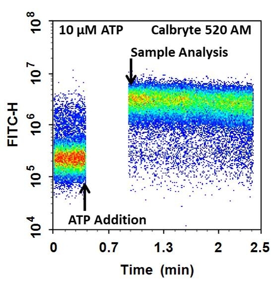
Cell Meter™ Flow Cytometric Calcium Assay Kit provides fluorescence-based assays for detecting intracellular calcium mobilization using a flow cytometer. It can be used for kinetic reading or for endpoint reading. After loading the Calbryte™ 520 AM dye into cells of interest, simply wash the cells and add the calcium flux agonist, one can then read the sample via a flow cytometer using kinetic reading mode or endpoint reading mode. Calbryte™ 520 AM can cross cell membrane passively by diffusion. Once inside the cells, the lipophilic blocking groups of Calbryte™ 520 AM are cleaved by esterase, resulting in a negatively charged fluorescent dye that stays inside cells. Its fluorescence is greatly enhanced upon binding to calcium. When cells expressing GPCR of interest are stimulated with an agonist, the receptor signals the release of intracellular calcium, which significantly increases the fluorescence of Calbryte™ 520. The Cell Meter™ Flow Cytometric Calcium Assay Kit can be used for monitoring cellular calcium flux as well as cell sorting.
Platform
Flow cytometer
Excitation | 488 nm laser |
Emission | 530/30 nm filter |
Instrument specification(s) | FITC channel |
Other instruments
ArrayScan, FDSS, FlexStation, IN Cell Analyzer, NOVOStar, ViewLuxComponents
Example protocol
AT A GLANCE
Protocol summary
- Prepare cells in Assay Buffer
- Add Calbryte™ 520 AM dye-loading solution (1 µL)
- Incubate at 37°C for 30 minutes
- Wash the cells
- Add calcium flux stimulator
- Monitor fluorescence intensity with flow cytometer using 530/30 nm filter (FITC channel)
Important notes
Thaw all the kit components at room temperature before starting the experiment.
PREPARATION OF STOCK SOLUTION
Unless otherwise noted, all unused stock solutions should be divided into single-use aliquots and stored at -20 °C after preparation. Avoid repeated freeze-thaw cycles.
Calbryte™ 520 AM stock solution (500X):
Add 100 µL of DMSO (Not provided) into the vial of Calbryte™ 520 AM stock solution (Component A) and mix them well. Note: 100 µL of Calbryte™ 520 AM stock solution is enough for 100 assays. Unused Calbryte™ 520 AM stock solution can be aliquoted and stored at < -20 °C for more than one month if the tubes are sealed tightly. Protect from light and avoid repeated freeze-thaw cycles.
SAMPLE EXPERIMENTAL PROTOCOL
- Remove cell culture medium and add 0.5 mL of Assay Buffer. Note: For adherent cells and non-adherent cells, 4 X10⁵ – 8 X10⁵ and 1X10⁶ - 2X10⁶ are recommended to use, respectively. Each cell line should be evaluated on the individual basis to determine the optimal cell density for the intracellular calcium mobilization.
- Add 1 µL Calbryte™ 520 AM stock solution (500X) into 0.5 mL non-adherent or adherent cells in Assay Buffer (Component B). Note: If your cells (such as CHO cells) contain organic anion-transports, then probenecid (Component C) may be added to the dye working solution (final in well concentration will be 0.125-1 mM) to reduce leakage of the de-esterified indicators.
- Incubate the cells at 37°C for 30 minutes.
- For non-adherent cells, centrifuge the cells and remove the dye. Re-suspend the cells in 0.4 mL HHBS (Component D). For adherent cells, use 0.5 mM EDTA to gently lift the cells from the plate and centrifuge. Re-suspend the cells in 0.4 mL HHBS (Component D). Note: For detaching adherent cells from the plate, enzymatic reagents (e.g. trypsin, Accutase) can be considered but need to be tested to make sure the receptor of interest on the cell surface is not affected.
- Prepare 5X agonist compound with HHBS or your desired buffer.
- Analyze the sample before and after the addition of 100 µL of the prepared agonist on a flow cytometer using 530/30 nm filter (FITC channel). Note: To achieve the best results, it is important to run the assay within 1 minute after the addition of the agonist. It is also important to make sure the time between the agonist addition and the beginning of the actual reading stays constant for all the samples.
Spectrum
Open in Advanced Spectrum Viewer


Spectral properties
Excitation (nm) | 493 |
Emission (nm) | 515 |
Quantum yield | 0.751 |
Images

Figure 1. The ATP induced intracellular calcium release was measured by Cell Meter™ Flow Cytometric Calcium Assay Kit in CHO-K1 cells. Cells were incubated with Calbryte™ 520 AM dye for 30 min at 37 °C before 10 µM ATP was added into the cells. The baseline was acquired and the rest of the cells were analyzed after the addition of ATP. The response was measured over time. The analysis was done on NovoCyte™ 3000 Flow Cytometer. The arrows on the graph indicate the time (30 sec) between addition of ATP and the actual analysis.

Figure 2. The ATP dose dependent intracellular calcium release was measured by Cell Meter™ Flow Cytometric Calcium Assay Kit in CHO-K1 cells. Cells were incubated with Calbryte™ 520 AM dye for 30 min at 37 °C before ATP was added into the cells. The baseline was acquired and the rest of the cells were analyzed after the addition of ATP. The response was measured over time. The analysis was done on NovoCyte™ 3000 Flow Cytometer. A. 10 µM, 1 µM or 0 µM ATP were added to the cells. The arrows on the graph indicate the time (30 sec) between addition of ATP and the actual analysis. B. Time-dependent changes of fluorescent signal.
Citations
View all 2 citations: Citation Explorer
iPLA2 Inhibition Blocks LysoPC-Induced TRPC6 Externalization and Promotes Re-Endothelialization of Carotid Injuries in Hypercholesterolemic Mice
Authors: Putta, Priya and Chaudhuri, Pinaki and Guardia-Wolff, Rocio and Rosenbaum, Michael A and Graham, Linda M
Journal: Cell Calcium (2023): 102734
Authors: Putta, Priya and Chaudhuri, Pinaki and Guardia-Wolff, Rocio and Rosenbaum, Michael A and Graham, Linda M
Journal: Cell Calcium (2023): 102734
Inhibition of P110$\alpha$ and P110$\delta$ catalytic subunits of PI3 kinase reverses impaired arterial healing after injury in hypercholesterolemic male mice
Authors: Chaudhuri, Pinaki and Smith, Andrew H and Graham, Linda M and Rosenbaum, Michael A
Journal: American Journal of Physiology-Cell Physiology (2021): C943--C955
Authors: Chaudhuri, Pinaki and Smith, Andrew H and Graham, Linda M and Rosenbaum, Michael A
Journal: American Journal of Physiology-Cell Physiology (2021): C943--C955
References
View all 34 references: Citation Explorer
Fluorescence absorbance inner-filter decomposition: the role of emission shape on estimates of free Ca(2+) using Rhod-2
Authors: Territo PR, Heil J, Bose S, Evans FJ, Balaban RS.
Journal: Appl Spectrosc (2007): 138
Authors: Territo PR, Heil J, Bose S, Evans FJ, Balaban RS.
Journal: Appl Spectrosc (2007): 138
Protein kinase C and myocardial calcium handling during ischemia and reperfusion: lessons learned using Rhod-2 spectrofluorometry
Authors: Stamm C, del Nido PJ.
Journal: Thorac Cardiovasc Surg (2004): 127
Authors: Stamm C, del Nido PJ.
Journal: Thorac Cardiovasc Surg (2004): 127
Novel fluo-4 analogs for fluorescent calcium measurements
Authors: Martin VV, Beierlein M, Morgan JL, Rothe A, Gee KR.
Journal: Cell Calcium (2004): 509
Authors: Martin VV, Beierlein M, Morgan JL, Rothe A, Gee KR.
Journal: Cell Calcium (2004): 509
Kinetic characterization of novel NR2B antagonists using fluorescence detection of calcium flux
Authors: Bednar B, Cunningham ME, Kiss L, Cheng G, McCauley JA, Liverton NJ, Koblan KS.
Journal: J Neurosci Methods (2004): 247
Authors: Bednar B, Cunningham ME, Kiss L, Cheng G, McCauley JA, Liverton NJ, Koblan KS.
Journal: J Neurosci Methods (2004): 247
Cytosolic calcium in the ischemic rabbit heart: assessment by pH- and temperature-adjusted rhod-2 spectrofluorometry
Authors: Stamm C, Friehs I, Choi YH, Zurakowski D, McGowan FX, del Nido PJ.
Journal: Cardiovasc Res (2003): 695
Authors: Stamm C, Friehs I, Choi YH, Zurakowski D, McGowan FX, del Nido PJ.
Journal: Cardiovasc Res (2003): 695
Calcium measurements in perfused mouse heart: quantitating fluorescence and absorbance of Rhod-2 by application of photon migration theory
Authors: Du C, MacGowan GA, Farkas DL, Koretsky AP.
Journal: Biophys J (2001): 549
Authors: Du C, MacGowan GA, Farkas DL, Koretsky AP.
Journal: Biophys J (2001): 549
Calibration of the calcium dissociation constant of Rhod(2)in the perfused mouse heart using manganese quenching
Authors: Du C, MacGowan GA, Farkas DL, Koretsky AP.
Journal: Cell Calcium (2001): 217
Authors: Du C, MacGowan GA, Farkas DL, Koretsky AP.
Journal: Cell Calcium (2001): 217
Changes in mitochondrial Ca2+ detected with Rhod-2 in single frog and mouse skeletal muscle fibres during and after repeated tetanic contractions
Authors: Lannergren J, Westerblad H, Bruton JD.
Journal: J Muscle Res Cell Motil (2001): 265
Authors: Lannergren J, Westerblad H, Bruton JD.
Journal: J Muscle Res Cell Motil (2001): 265
Rhod-2 based measurements of intracellular calcium in the perfused mouse heart: cellular and subcellular localization and response to positive inotropy
Authors: MacGowan GA, Du C, Glonty V, Suhan JP, Koretsky AP, Farkas DL.
Journal: J Biomed Opt (2001): 23
Authors: MacGowan GA, Du C, Glonty V, Suhan JP, Koretsky AP, Farkas DL.
Journal: J Biomed Opt (2001): 23
Mitochondrial free calcium levels (Rhod-2 fluorescence) and ultrastructural alterations in neuronally differentiated PC12 cells during ceramide-dependent cell death
Authors: Muriel MP, Lambeng N, Darios F, Michel PP, Hirsch EC, Agid Y, Ruberg M.
Journal: J Comp Neurol (2000): 297
Authors: Muriel MP, Lambeng N, Darios F, Michel PP, Hirsch EC, Agid Y, Ruberg M.
Journal: J Comp Neurol (2000): 297
Application notes
A Comparison of Fluorescent Red Calcium Indicators for Detecting Intracellular Calcium Mobilization in CHO Cells
A Meta-Analysis of Common Calcium Indicators
A New Red Fluorescent & Robust Screen Quest™ Rhod-4™ Ca2+Indicator for Screening GPCR & Ca2+ Channel Targets
A New Robust No-Wash FLIPR Calcium Assay Kit for Screening GPCR and Calcium Channel Targets
A Novel NO Wash Probeniceid-Free Calcium Assay for Functional Analysis of GPCR and Calcium Channel Targets
FAQ
Do you offer any products for measuring intracellular calcium concentration or movement by flow cytometry?
Are there any calcium indicators that don't require probenecid (PBC)?
Are there upgraded trypan blue derivatives for cell viability testing?
Can I intracellularly measure mitochondria calcium flux and changes in mitochondria membrane potential at the same time?
Does EDTA inactivate proteinase K?
Are there any calcium indicators that don't require probenecid (PBC)?
Are there upgraded trypan blue derivatives for cell viability testing?
Can I intracellularly measure mitochondria calcium flux and changes in mitochondria membrane potential at the same time?
Does EDTA inactivate proteinase K?