Products
Services
Resources
Selection Guides
About
Cyanine 5 monosuccinimidyl ester
equivalent to Cy5® NHS ester
Cy5 NHS Ester is a far-red fluorescent labeling reagent that efficiently conjugates to amine groups on proteins and peptides, providing excellent tissue penetration and minimal background for in vivo imaging applications.
- Far-red fluorescence: Deep tissue penetration with minimal autofluorescence for in vivo imaging applications
- 633 nm laser compatible: Optimized for standard red laser lines in confocal microscopy and flow cytometry
- High-efficiency protein labeling: NHS ester chemistry provides 80–95% conjugation yields with antibodies and proteins
- Alternative to Alexa Fluor 647: Spectrally equivalent with improved cost-effectiveness for large-scale labeling projects
| Catalog | Size | Price | Quantity |
|---|---|---|---|
| 151 | 1 mg | Price |

Physical properties
| Molecular weight | 855.07 |
| Solvent | DMSO |
Spectral properties
| Correction factor (260 nm) | 0.02 |
| Correction factor (280 nm) | 0.03 |
| Correction factor (482 nm) | 0.009 |
| Correction factor (565 nm) | 0.09 |
| Extinction coefficient (cm -1 M -1) | 250000 1 |
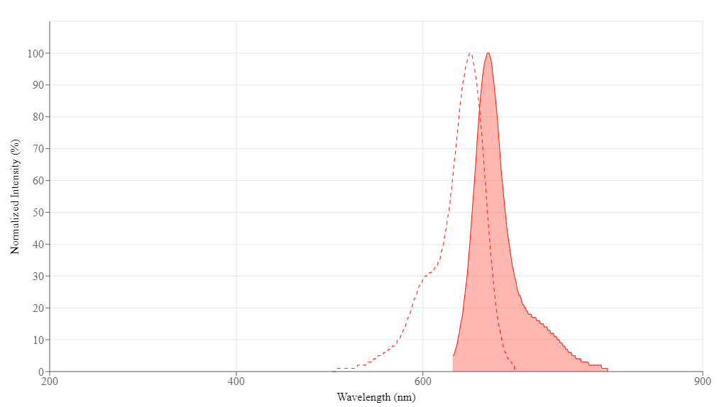
| Excitation (nm) | 651 |
| Emission (nm) | 670 |
| Quantum yield | 0.27 1 , 0.42 |
Storage, safety and handling
| H-phrase | H303, H313, H333 |
| Hazard symbol | XN |
| Intended use | Research Use Only (RUO) |
| R-phrase | R20, R21, R22 |
| Storage | Freeze (< -15 °C); Minimize light exposure |
| UNSPSC | 12171501 |
| CAS | 146368-14-1 |
Contact us
| Telephone | |
| Fax | |
| sales@aatbio.com | |
| International | See distributors |
| Bulk request | Inquire |
| Custom size | Inquire |
| Technical Support | Contact us |
| Request quotation | Request |
| Purchase order | Send to sales@aatbio.com |
| Shipping | Standard overnight for United States, inquire for international |
Page updated on September 27, 2024