Products
Services
Resources
Selection Guides
About
Cyanine 7 monosuccinimidyl ester
equivalent to Cy7® NHS ester
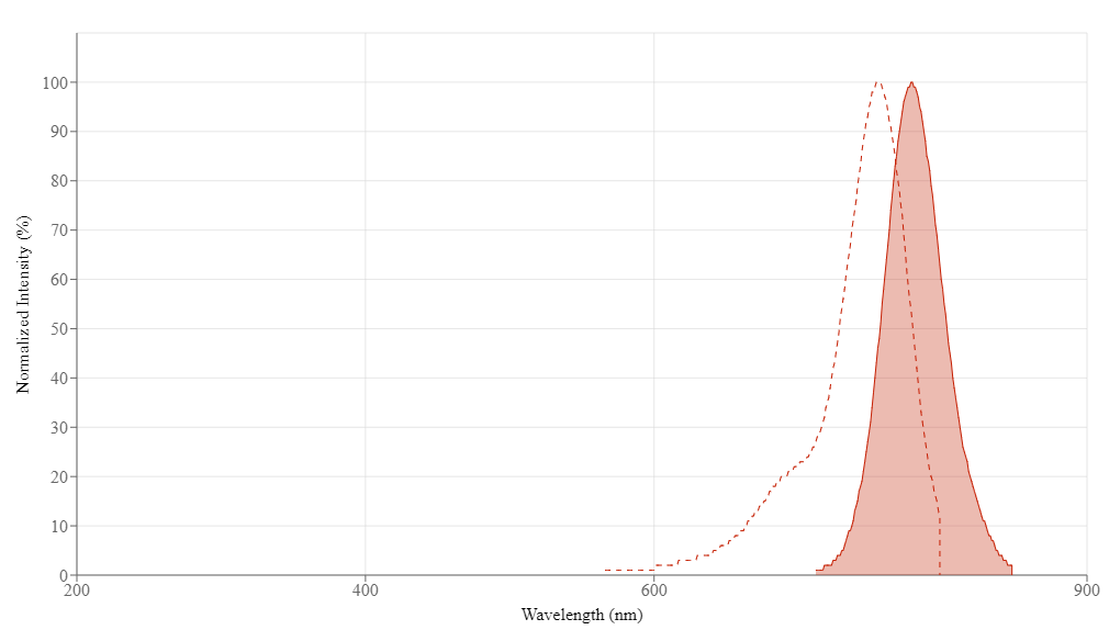
Cy7 NHS Ester is a near-infrared fluorescent dye (Ex/Em: 756/779 nm) for amine-reactive labeling that enables deep tissue imaging and whole-animal fluorescence detection with minimal autofluorescence interference.
- Near-Infrared Emission (756/779 nm): Enables whole-animal imaging with minimal tissue absorption and background
- Multimodal Imaging Compatibility: Suitable for fluorescence tomography, surgical guidance, and lymph node mapping
- Enhanced Aqueous Solubility: Triethylammonium salt formulation improves reconstitution compared to potassium salts
| Catalog | Size | Price | Quantity |
|---|---|---|---|
| 161 | 1 mg | Price |

Physical properties
| Molecular weight | 881.11 |
| Solvent | DMSO |
Spectral properties
| Correction factor (260 nm) | 0.05 |
| Correction factor (280 nm) | 0.036 |
| Correction factor (482 nm) | 0.0005 |
| Correction factor (565 nm) | 0.0193 |
| Correction factor (650 nm) | 0.165 |
| Extinction coefficient (cm -1 M -1) | 250000 |
| Excitation (nm) | 756 |
| Emission (nm) | 779 |
| Quantum yield | 0.3 |
Usage and storage
| Intended use | Research Use Only (RUO) |
| Storage | Freeze (< -15 °C); Minimize light exposure |
| CAS | 477908-53-5 |
Contact us
| Telephone | |
| Fax | |
| sales@aatbio.com | |
| International | See distributors |
| Bulk request | Inquire |
| Custom size | Inquire |
| Technical Support | Contact us |
| Request quotation | Request |
| Purchase order | Send to sales@aatbio.com |
| Shipping | Standard overnight for United States, inquire for international |
Page updated on September 11, 2024