Products
Services
Resources
Selection Guides
About
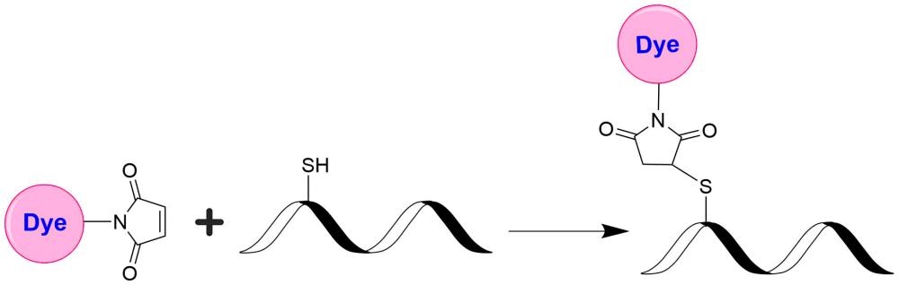
iFluor® 770 maleimide
AAT Bioquest's iFluor® dyes are optimized for labeling proteins, particularly antibodies. These dyes are bright, photostable, and have minimal quenching on proteins. They can be well excited by the major laser lines of fluorescence instruments (e.g., 350, 355, 405, 488, 555, 633, 638, 647, 660, and 802 nm). iFluor® 770 dyes have fluorescence excitation and emission maxima of ~777 nm and ~797 nm respectively. These spectral characteristics make them a unique color for fluorescence imaging and flow cytometry applications. iFluor® 770 is an excellent acceptor dye for preparing tandem colors with APC and PE. These iFluor® 770 tandem colors offer a set of unique color profiles for spectral flow cytometry. iFluor® 770 is one of the brightest NIR dyes, and some of its antibody conjugates are significantly brighter than those prepared with IRDyes of similar wavelengths, such as IRDye 800RS. iFluor® 770 maleimide is a thiol-reactive form used to conjugate with thiol-containing molecules such as reduced antibodies, thiol-modified oligos, and peptides.
| Catalog | Size | Price | Quantity |
|---|---|---|---|
| 1370 | 1 mg | Price |

Physical properties
| Solvent | DMSO |
Spectral properties
| Absorbance (nm) | 772 |
| Correction factor (260 nm) | 0.09 |
| Correction factor (280 nm) | 0.08 |
| Extinction coefficient (cm -1 M -1) | 250000 1 |
| Excitation (nm) | 777 |
| Emission (nm) | 797 |
| Quantum yield | 0.16 |
Storage, safety and handling
| H-phrase | H303, H313, H333 |
| Hazard symbol | XN |
| Intended use | Research Use Only (RUO) |
| R-phrase | R20, R21, R22 |
| Storage | Freeze (< -15 °C); Minimize light exposure |
| UNSPSC | 12171501 |
Contact us
| Telephone | |
| Fax | |
| sales@aatbio.com | |
| International | See distributors |
| Bulk request | Inquire |
| Custom size | Inquire |
| Technical Support | Contact us |
| Request quotation | Request |
| Purchase order | Send to sales@aatbio.com |
| Shipping | Standard overnight for United States, inquire for international |
Page updated on May 24, 2026