logo
AAT Bioquest

PhosphoWorks™ Fluorimetric ADP Assay Kit *Red Fluorescence*

ADP is involved in many biological reactions such as protein kinases. Our ADP assay kit can be used for assaying protein kinase reactions universally by monitoring ADP formation, which is directly proportional to enzyme phosphotransferase activity and is measured fluorimetrically. This kit provides a fast, simple and homogeneous assay for measuring ADP formation or depletion. The assay is continuous, and can be easily adapted to automation. The kit is convenient, requiring minimal hands-on time. Protein kinases are of interest to researchers involved in drug discovery due to their broad relevance to diseases such as cancer and other proliferative diseases, inflammatory diseases, metabolic disorders and neurological diseases. Most of commercial protein kinase assay kits are either based on monitoring of phosphopeptide formation or ATP deletion. Our ADP assay kit is more robust for assaying protein kinases than most of commercial kinase assay kits.
Example protocol

AT A GLANCE

Protocol summary

  1. Run kinase reaction (20 µL)
  2. Add ADP Sensor Buffer (20 µL)
  3. Add ADP Sensor (10 µL)
  4. Incubate at room temperature for 15 minutes - 1 hour
  5. Monitor fluorescence intensity

Important notes
Thaw all the six components at room temperature before use. Avoid direct exposure of ADP Sensor I (Component B1) to light. 

PREPARATION OF STOCK SOLUTION

Unless otherwise noted, all unused stock solutions should be divided into single-use aliquots and stored at -20 °C after preparation. Avoid repeated freeze-thaw cycles.

1. ADP Sensor I stock solution (50X):
Add 20 µL DMSO (Component B3) into vial of ADP Sensor I (Component B1).

2. ADP Sensor stock solution (1X):
Add 20 µL of 50X ADP Sensor I stock solution into vial of ADP Sensor II (Component B2).

3. ADP standard solution (300 mM):
Add 100 µL of H2O into ADP Standard (Component C) to make a 300 mM ADP stock solution.

PREPARATION OF STANDARD SOLUTION

ADP standard

For convenience, use the Serial Dilution Planner: https://www.aatbio.com/tools/serial-dilution/21655

Make serial dilutions of ADP standard in the kinase reaction buffer by including a sample without ADP for measuring background fluorescence. Note: Typically, ADP concentrations ranging from 0.05 to 30 µM are appropriate.

SAMPLE EXPERIMENTAL PROTOCOL

Table 1. Layout of ADP standards and test samples in a solid black 96-well microplate. SD = ADP standard (SD1 - SD7, 0.05 to 30 uM); BL = blank control; TS = test sample.

BLBLTSTS
SD1SD1......
SD2SD2......
SD3SD3  
SD4SD4  
SD5SD5  
SD6SD6  
SD7SD7  

Table 2. Reagent composition for each well.

WellVolumeReagent
SD1 - SD720 µLserial dilution (0.05 to 30 µM)
BL20 µLADP assay buffer
TS20 µLkinase reaction


Run kinase reaction (Reagents are not provided for this step):

  1. Prepare 20 µL of kinase reaction solution as desired. The components of kinase reaction should be optimized as needed (e.g., an optimized buffer system might be required for a specific kinase reaction). In most cases, ADP assay buffer (Component D) can also be used to run kinase reaction if you do not have the optimized kinase buffer.

  2. The Amplite™ Fluorimetric ADP Assay Kit is used to determine the ADP formation. The ADP Sensor is unstable in the presence of thiols such as DTT and β-mercaptoethanol. Final thiol concentration higher than 10 µM would significantly decrease the assay dynamic range.

Run Amplite™ ADP assay:

  1. Add 20 µL of ADP Sensor Buffer (Component A) and 10 µL of ADP Sensor stock solution into each well filled with the 20 µL kinase reaction solution to make the total ADP assay volume of 50 µL/well. Note: The ADP Sensor is unstable in the presence of thiols such as DTT and β-mercaptoethanol. Final thiol concentration higher than 10 µM would significantly decrease the assay dynamic range. Note: The ADP assay should be run at pH from 6.5 to 7.4.

  2. Incubate the reaction mixture at room temperature for 15 minutes to 1 hour.

  3. Monitor the fluorescence intensity with a fluorescence plate reader at Ex/Em = 540/590 nm.

Citations

View all 4 citations: Citation Explorer
Discovery of Non-ATP-Competitive Inhibitors of Polo-like Kinase 1
Authors: Yun, Taikangxiang and Qin, Tan and Liu, Ying and Lai, Luhua
Journal: ChemMedChem (2016): 713--717
Cell polarity kinase MST4 cooperates with cAMP-dependent kinase to orchestrate histamine-stimulated acid secretion in gastric parietal cells
Authors: Jiang, Hao and Wang, Wenwen and Zhang, Yin and Yao, William W and Jiang, Jiying and Qin, Bo and Yao, Wendy Y and Liu, Fusheng and Wu, Huihui and Ward, Tarsha L and others, undefined
Journal: Journal of Biological Chemistry (2015): 28272--28285
Regulation of NDR1 activity by PLK1 ensures proper spindle orientation in mitosis
Authors: Yan, Maomao and Chu, Lingluo and Qin, Bo and Wang, Zhikai and Liu, Xing and Jin, Changjiang and Zhang, Guanglan and Gomez, Marta and Hergovich, Alex and er , undefined and Chen, Zhengjun and others, undefined
Journal: Scientific reports (2015): 10449
Trypanosoma brucei vacuolar transporter chaperone 4 (TbVtc4) is an acidocalcisome polyphosphate kinase required for in vivo infection
Authors: L, undefined and er, Noelia and Ulrich, Paul N and Docampo, Roberto
Journal: Journal of Biological Chemistry (2013): 34205--34216

References

View all 36 references: Citation Explorer
The ADP/ATP ratio: A novel predictive assay for quality assessment of isolated pancreatic islets
Authors: Goto M, Holgersson J, Kumagai-Braesch M, Korsgren O.
Journal: Am J Transplant (2006): 2483
Negative correlation between poly-ADP-ribosylation of spleen cell histone proteins and initial duration of dimethylnitrosamine exposure to mice in vivo measured by Western blot immunoprobe assay: a possible biomarker for cancer detection
Authors: Devi BJ, Schneeweiss FH, Sharan RN.
Journal: Cancer Detect Prev (2005): 66
A novel fluorometric assay for ADP-ribose pyrophosphatase activity
Authors: Song EK, Park HJ, Kim JS, Lee HH, Kim UH, Han MK.
Journal: J Biochem Biophys Methods (2005): 161
An assay for adenosine 5'-diphosphate (ADP)-glucose pyrophosphorylase that measures the synthesis of radioactive ADP-glucose with glycogen synthase
Authors: Yep A, Bejar CM, Ballicora MA, Dubay JR, Iglesias AA, Preiss J.
Journal: Anal Biochem (2004): 52
A nonradiometric, high-throughput assay for poly(ADP-ribose) glycohydrolase (PARG): application to inhibitor identification and evaluation
Authors: Putt KS, Hergenrother PJ.
Journal: Anal Biochem (2004): 256
Page updated on December 11, 2024

Ordering information

Price
Unit size
Catalog Number21655
Quantity
Add to cart

Additional ordering information

Telephone1-800-990-8053
Fax1-800-609-2943
Emailsales@aatbio.com
InternationalSee distributors
Bulk requestInquire
Custom sizeInquire
Technical SupportContact us
Purchase orderSend to sales@aatbio.com
ShippingStandard overnight for United States, inquire for international
Request quotation

Storage, safety and handling

H-phraseH303, H313, H333
Hazard symbolXN
Intended useResearch Use Only (RUO)
R-phraseR20, R21, R22
UNSPSC12352200

Platform

Fluorescence microplate reader

Excitation540 nm
Emission590 nm
Cutoff570 nm
Recommended plateSolid black

Components

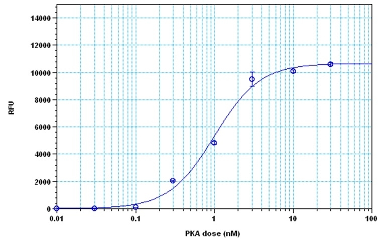
The detection of protein kinase A with PhosphoWorks™ Fluorimetric ADP Assay Kit. The kinase was incubated in the presence of ATP and kemptide peptide substrate for 30 minutes, and ADP generation was detected after 30 minutes incubation using the Amplite® Fluorimetric Kinase Assay Kit.