logo
AAT Bioquest

Portelite™ Fluorimetric DNA Quantitation Kit with Broad Dynamic Range *Optimized for Cytocite™ and Qubit™ Fluorometers*

OverviewpdfSDSpdfProtocol


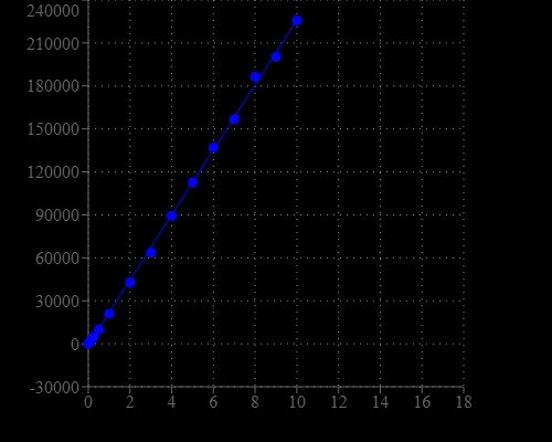
DNA Quantitation is a very important task in DNA sample preparations for various analyses. Portelite™ Fluorimetric DNA Quantitation Kit provides a rapid method to quantify dsDNA with Helixyte™ Green BR using a hand-held fluorometer. It is optimized for Cytocite™ and Qubit™ fluorometers. Portelite™ Fluorimetric DNA Quantitation assay is linear over three orders of magnitude. The assay is highly selective for double-stranded DNA (dsDNA) over RNA and is optimized for measuring dsDNA concentrations from 10 pg/µL to 10 ng/µL. Helixyte™ Green BR exhibits large fluorescence enhancement upon binding to dsDNA, and it is a few magnitudes more sensitive than UV absorbance readings.

Platform


Qubit Fluorometer

Excitation480 nm
Emission530 nm
Instrument specification(s)0.2 mL PCR vial

CytoCite Fluorometer

Excitation480 nm
Emission530 nm
Instrument specification(s)0.2 mL PCR vial

Components


Example protocol


AT A GLANCE

Protocol summary

  1. Prepare Helixyte™ Green BR working solution
  2. Add 190 uL 1X Helixyte™ Green BR working solution into each 0.2 mL PCR tube
  3. Add 10 uL DNA standards or test samples into each tube
  4. Incubate at room temperature for 2 minutes
  5. Monitor fluorescence with CytoCite™ fluorometer or Qubit™

Important notes
Bring all the components at room temperature before opening.

PREPARATION OF WORKING SOLUTION

Helixyte™ Green BR working solution:
Make a 200-fold dilution of Portelite™ dsDNA reagent (Component A) in DNA Assay Buffer (Component B). For example, to prepare enough working solution for 8 samples, add 5 uL of Helixyte™ Green BR (Component A) into 1 mL of DNA Assay Buffer (Component B). Protect the working solution from light by covering it with foil or placing it in the dark. Note: Protect the working solution from light by covering it with foil or placing it in the dark. We recommend preparing this solution in a plastic container rather than glass, as the dye may adsorb to glass surfaces.  For best results, this solution should be used within a few hours of its preparation.

SAMPLE EXPERIMENTAL PROTOCOL

The acceptable sample volume could be a range from 1~20 uL depending on the estimate concentration of DNA sample. The recommend sample volume is 10 uL with the DNA concentration in 0.2~100 ng/uL range. If other sample volume is being used, please adjust the dilution factor in the concentration calculations. 

 The following protocol is generated based on 10 uL sample volume with the DNA concentration in 0.2~100 ng/uL range.

  1. Add 190 uL 1X Helixyte™ Green BR working solution into each Cytocite™ sample tube (#CCT100) or equivalent 0.2 mL PCR tube. Note: Use thin-wall, polypropylene, clear 0.2 mL PCR tubes such as#CCT100.

  2. Add DNA standards or test samples 10 uL into each tube, and then mix by vortexing 2~3 seconds.

  3. Incubate all tubes at room temperature for 2 minutes.

  4. Insert the samples into CytoCite™ or Quibit™ and monitor the fluorescence with green fluorescence channel. Follow the procedure appropriate for CytoCite™ Fluorometer. See the link below for detailed instructions: https://devices.aatbio.com/documentation/user-manual-for-cytocite-fluorometer

PREPARATION OF STANDARD CALIBRATION CURVE

For Portelite™ assays, you have the choice to make a calibration curve with the DNA standards. Here is a brief protocol to generate a customized DNA standard curve:

  1. Perform 1/3 serial dilution with 100 ng/uL with DNA Standard BR #2 (Component D) in DNA Assay Buffer (Component B) to get 30, 10, 3, 1, 0.3, 0.1 and 0 ng/uL DNA standard dilutions.

  2. Add 190 uL of Helixyte™ Green BR working solution into each tube.

  3. Add 10 uL standards or 10 uL samples into a 0.2 mL PCR tube.

  4. Incubate the reaction at room temperature for 2 minutes.

  5. Insert the samples into CytoCite™ and monitor the fluorescence with green fluorescence channel.

Images


Citations


View all 32 citations: Citation Explorer
A new reporter design based on DNA origami nanostructures for quantification of short oligonucleotides using microbeads
Authors: Choi, Y., Schmidt, C., Tinnefeld, P., Bald, I., Rodiger, S.
Journal: Sci Rep (2019): 4769
Flat-top TIRF illumination boosts DNA-PAINT imaging and quantification
Authors: Stehr, F., Stein, J., Schueder, F., Schwille, P., Jungmann, R.
Journal: Nat Commun (2019): 1268
Effects of Quantification Methods, Isolation Kits, Plasma Biobanking, and Hemolysis on Cell-Free DNA Analysis in Plasma
Authors: Streleckiene, G., Forster, M., Inciuraite, R., Lukosevicius, R., Skieceviciene, J.
Journal: Biopreserv Biobank (2019): ersion="1.0" encoding="UTF-8" ?>17645.enlEndN
A universal fluorescence-based toolkit for real-time quantification of DNA and RNA nuclease activity
Authors: Sheppard, E. C., Rogers, S., Harmer, N. J., Chahwan, R.
Journal: Sci Rep (2019): 8853
Molecular-Recognition-Based DNA Nanodevices for Enhancing the Direct Visualization and Quantification of Single Vesicles of Tumor Exosomes in Plasma Microsamples
Authors: He, D., Ho, S. L., Chan, H. N., Wang, H., Hai, L., He, X., Wang, K., Li, H. W.
Journal: Anal Chem (2019): 2768-2775
Quantification of fixed adherent cells using a strong enhancer of the fluorescence of DNA dyes
Authors: Ligasova, A., Koberna, K.
Journal: Sci Rep (2019): 8701
Identification and Quantification of Heterogeneously-methylated DNA Fragments Using Epiallele-sensitive Droplet Digital Polymerase Chain Reaction (EAST-ddPCR)
Authors: Menschikowski, M., J and eck, C., Friedemann, M., Richter, S., Thiem, D., Lange, B. S., Suttorp, M.
Journal: Cancer Genomics Proteomics (2018): 299-312
A fluorescent reporter for quantification and enrichment of DNA editing by APOBEC-Cas9 or cleavage by Cas9 in living cells
Authors: St Martin, A., Salamango, D., Serebrenik, A., Shaban, N., Brown, W. L., Donati, F., Munagala, U., Conticello, S. G., Harris, R. S.
Journal: Nucleic Acids Res (2018): e84
Three-Dimensional FRET Multiplexing for DNA Quantification with Attomolar Detection Limits
Authors: Qiu, X., Guo, J., Xu, J., Hildebr and t, N.
Journal: J Phys Chem Lett (2018): 4379-4384
Quantification of plasmid DNA standards for U.S. EPA fecal indicator bacteria qPCR methods by droplet digital PCR analysis
Authors: Sivaganesan, M., Varma, M., Siefring, S., Haugl and , R.
Journal: J Microbiol Methods (2018): 135-142