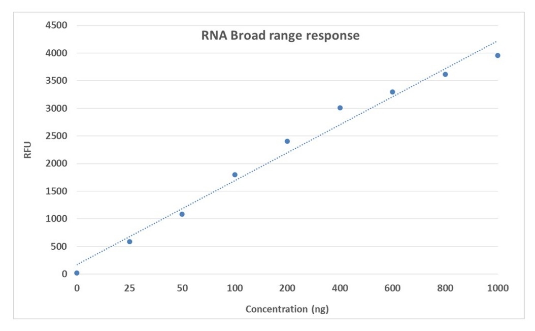
Portelite™ Fluorimetric RNA Quantification Kit *20-1000 ng Broad Range*
Product key features
Portelite™ Fluorimetric RNA Quantification Assay Kit accurately measures RNA across a broad range for research applications.
- Broad range detection: Measures RNA in the 20–1000 ng range with high accuracy using a fluorescence-based assay
- High specificity: Selectively detects RNA with minimal interference from DNA, proteins, or other contaminants
- Applications: Suitable for RT-qPCR, RNA-seq, cDNA synthesis, and other RNA-based studies
- Comparable alternative: Provides a performance advantage over traditional absorbance-based RNA quantification kits with reduced interference and higher accuracy
Product description
The Portelite™ Fluorimetric RNA Quantification Assay Kit provides a reliable and efficient method for measuring RNA across a broad concentration range. Using a proprietary RNA-selective fluorescent dye, this assay offers superior accuracy and specificity compared to traditional absorbance-based methods, minimizing interference from DNA, proteins, and other non-RNA components.
This kit is compatible with a variety of RNA sample types, including total RNA and mRNA. Its streamlined protocol reduces variability and speeds up RNA analysis, making it well-suited for routine use. The Portelite™ Fluorimetric RNA Quantification Assay Kit is ideal for downstream applications such as RT-qPCR, RNA-seq, or cDNA synthesis, where RNA measurement precision is critical.
Example protocol
AT A GLANCE
- Add StrandBrite™ Red RNA BR reagent working solution (200 µL).
- Add test samples (10 µL).
- Incubate at room temperature for 5 minutes.
- Monitor fluorescence intensity in Qubit™ fluorometer using red filter.
Important: The following protocol is provided as an example for quantifying total RNA with StrandBrite™ Red RNA BR. Warm all the components to room temperature before opening. No data are available for the mutagenicity or toxicity of StrandBrite™ Red RNA BR stain. Because this reagent binds to nucleic acids, it should be treated as a potential mutagen and handled with appropriate care. The DMSO stock solution should be handled with particular caution as DMSO is known to facilitate the entry of organic molecules into tissues.
PREPARATION OF WORKING SOLUTION
Add 2.5 μL StrandBrite™ Red RNA BR (Component A) into 1 mL of RNA Assay Buffer (Component B) and mix well. Protect the working solution from light by covering it with foil or placing it in the dark.
Note: 1 mL of working solution is enough for 5 tests.
Note: We recommend preparing this solution in a plastic container rather than glass, as the dye may adsorb to glass surfaces. For best results, this solution should be used within a few hours of its preparation.
SAMPLE EXPERIMENTAL PROTOCOL
The acceptable sample volume could range from 1~20 µL depending on the estimated concentration of RNA sample. The recommended sample volume is 10 µL with the RNA concentration in 2~100 ng/µL range. If other sample volume is being used, please adjust the dilution factor in the concentration calculations.
The following protocol is generated based on 10 µL sample volume with the RNA concentration in 2-100 ng/µL range.
- Add 200 µL StrandBrite™ Red RNA BR working solution into each CytoCite™ sample tube (AAT cat# CCT100) or equivalent 0.2 mL PCR tube.
Note: Use thin-wall, polypropylene, clear 0.2 mL PCR tubes such as AAT cat# CCT100. - Add 10 µL RNA standard #1 and #2 or test samples into each tube, and then mix by vortexing 2~3 seconds.
- Incubate all tubes at room temperature for 2 minutes.
- Insert the samples into Qubit™ and monitor the fluorescence with red fluorescence channel.
For StrandBrite™ assays, you have the choice to make a calibration curve with the RNA standards. Here is a brief protocol to generate a customized RNA standard curve:
- Perform dilution of 2 mg/mL RNA standard (Component D) to 100, 80, 60, 40, 20, 10, 5, 2 ng/uL in RNA Assay Buffer (Component B).
- Add 200 µL StrandBrite™ Red DNA BR working solution into each tube.
- Add 10 µL RNA standards or test samples into each tube, and then mix by vortexing 2-3 seconds.
- Incubate the reaction at room temperature for 2 minutes.
- Insert the samples into Qubit™ and monitor the fluorescence with red fluorescence channel.
References
Authors: Tanaka, Ami and Matsubayashi, Keiji and Odajima, Takeshi and Sakata, Hidekatsu and Iida, Juri and Kai, Kazuhiro and Goto, Naoko and Satake, Masahiro
Journal: Transfusion (2024): 335-347
Authors: Dyer, Wayne B and Suzuki, Kazuo and Levert, Angelique and Starr, Mitchell and Lloyd, Andrew R and Zaunders, John J
Journal: Frontiers in immunology (2024): 1382711
Authors: Hosokawa, Kazuo and Ohmori, Hitoshi
Journal: Biochemical and biophysical research communications (2024): 151070
Authors: Qureshi, Huma and Duran, Ana Carrasco and Mahmood, Hassan and Sarwar, Zahida and Mahmood, Khalid and Midde, Krishna and Canchola, Jesse A and Parkin, Neil T and La Brot, Benjamin
Journal: Journal of viral hepatitis (2024): 156-160
Authors: Kang, Brian and Zhang, Jiayu and Schwoerer, Michael P and Nelson, Amy N and Schoeman, Emily and Guo, Andrew and Ploss, Alexander and Myhrvold, Cameron
Journal: bioRxiv : the preprint server for biology (2023)