Products
Services
Resources
Selection Guides
About
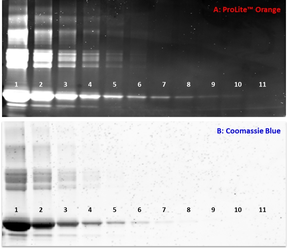
ProLite™ Orange Protein Gel Stain
5000X
Prolite™ Orange is an alternative protein stain that can be used to replace SYPRO Orange Protein Gel Stain (SYPRO is the trademark of ThermoFisher). Prolite™ Orange is a sensitive, ready-to-use fluorescent stain for total protein detection in 1D gels. The sensitivity of Prolite™ Orange is as good as or better than traditional silver staining techniques. Stained proteins can be viewed with a standard UV or blue-light transilluminator or imaging equipment containing the appropriate filters or lasers. Fluorescent stains are rapid, and highly sensitive for detecting total protein in protein electrophoresis gels and membranes. The Prolite™ Orange fluorescent stain can be used for total protein quantitation and can be viewed using a standard UV or blue-light transilluminator or with imaging instruments equipped with appropriate light sources.
| Catalog | Size | Price | Quantity |
|---|---|---|---|
| 18000 | 100 ul | Price | |
| 18001 | 1 ml | Price |

Physical properties
| Molecular weight | 486.72 |
| Solvent | Water |
Spectral properties
| Excitation (nm) | 484 |
| Emission (nm) | 586 |
Storage, safety and handling
| H-phrase | H303, H313, H333 |
| Hazard symbol | XN |
| Intended use | Research Use Only (RUO) |
| R-phrase | R20, R21, R22 |
| Storage | Freeze (< -15 °C); Minimize light exposure |
| UNSPSC | 12171501 |
Contact us
| Telephone | |
| Fax | |
| sales@aatbio.com | |
| International | See distributors |
| Bulk request | Inquire |
| Custom size | Inquire |
| Technical Support | Contact us |
| Request quotation | Request |
| Purchase order | Send to sales@aatbio.com |
| Shipping | Standard overnight for United States, inquire for international |
Page updated on June 18, 2026