Products
Services
Resources
Selection Guides
About
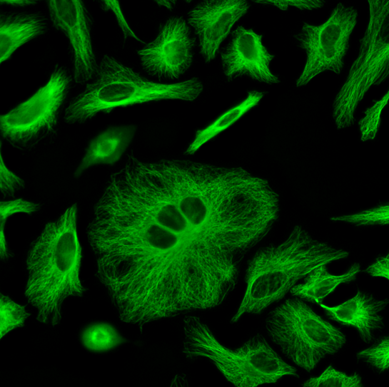
ReadiLink™ Rapid iFluor® 488 Antibody Labeling Kit
Production Scale
ReadiLink™ Rapid Antibody Labeling Kits, designed for production scale, provide a convenient and efficient method for labeling large volumes of antibodies with our superior iFluor® dyes, XFD dyes (equivalent to Alexa Fluor®), and various other labels. These kits utilize reactive fluorophores modified with succinimidyl ester (SE) functional groups, which selectively bind to primary amines on proteins, resulting in remarkably bright and photostable conjugates. Every kit contains all the necessary components for three distinct labeling reactions and features a user-friendly, pre-packed spin column for efficient dye removal, maximizing conjugate yield. Each vial of iFluor® 488 SE dye provided in the kit is precisely formulated to label 1 mg of purified protein or antibody. Before labeling, it is important to remove stabilizing proteins like BSA from the sample and refrain from using amine-rich buffers like Tris, which might disrupt the labeling process. iFluor® 488 SE is a bright green-fluorescent dye with excitation and emission maxima of ~491 nm and ~516 nm, making it an excellent alternative to FITC and Alexa Fluor® 488 (Alexa Fluor® is the trademark of Invitrogen). With ReadiLink™ Rapid Antibody Labeling kits, researchers can directly label primary antibodies, eliminating the need for secondary antibodies and enhancing panel-building flexibility.
| Catalog | Size | Price | Quantity |
|---|---|---|---|
| 5702 | 3 x 1 mg | Price |

Spectral properties
| Correction factor (260 nm) | 0.21 |
| Correction factor (280 nm) | 0.11 |
| Extinction coefficient (cm -1 M -1) | 75000 1 |
| Excitation (nm) | 491 |
| Emission (nm) | 516 |
| Quantum yield | 0.9 1 |
Usage and storage
| Intended use | Research Use Only (RUO) |
Contact us
| Telephone | |
| Fax | |
| sales@aatbio.com | |
| International | See distributors |
| Bulk request | Inquire |
| Custom size | Inquire |
| Technical Support | Contact us |
| Request quotation | Request |
| Purchase order | Send to sales@aatbio.com |
| Shipping | Standard overnight for United States, inquire for international |
Page updated on July 17, 2026