logo
AAT Bioquest

ReadiPrep™ Exosome Isolation Kit

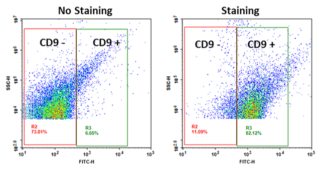
Exosomes from 100,000 HeLa cells (cultured for 16 hours in 10 mL serum-free medium) were isolated using ReadiPrep™ Exosome Isolation Kit (Cat# 60204) and detected using the Cell Navigator™ Flow Cytometric Exosome Staining Kit (Cat# 22426).
Ordering information
Price
Catalog Number
Unit Size
Quantity
Add to cart
Additional ordering information
Telephone1-800-990-8053
Fax1-800-609-2943
Emailsales@aatbio.com
InternationalSee distributors
Bulk requestInquire
Custom sizeInquire
ShippingStandard overnight for United States, inquire for international
Request quotation
Storage, safety and handling
H-phraseH303, H313, H333
Hazard symbolXN
Intended useResearch Use Only (RUO)
R-phraseR20, R21, R22
StorageRefrigerated (2-8 °C); Minimize light exposure
UNSPSC12352200

OverviewpdfSDSpdfProtocol


See also: Exosomes
Exosomes are nanoscale (30-150 nm), membrane-bound vesicles secreted by cells into the extracellular space, functioning as pivotal mediators in intercellular communication. They facilitate the transfer of proteins, lipids, and nucleic acids, including RNA species, between cells. This vesicular transport mechanism is implicated in various physiological and pathological processes, including immune modulation, disease progression, and genetic information exchange, making exosomes a focal point in both therapeutic and diagnostic research. The isolation of exosomes typically involves complex and time-consuming processes, such as ultracentrifugation, which can be challenging to execute. The ReadiPrep™ Exosome Isolation Kit offers a streamlined and effective approach to isolating intact exosomes from diverse biological samples, including cell culture media and serum. The principle behind this kit is based on the selective precipitation of exosomes by inducing the removal of water molecules from the solution. Following a brief, low-speed centrifugation step, the exosomes are precipitated and can subsequently be isolated through standard centrifugation. The isolated exosomal pellet is then resuspended in PBS or an equivalent buffer, rendering the exosomes suitable for further downstream analyses or purification via affinity-based methods.

Example protocol


SAMPLE EXPERIMENTAL PROTOCOL

Important Note

To ensure isolated exosomes originate from the cells, culture cells with exosome-depleted fetal bovine serum (FBS) or no FBS. Normal FBS contains a significantly higher amount of exosomes and may give false positive results. It is important to note that this kit has been tested only for exosome isolation from cell culture media.

Preparation of Cell Samples
  1. Grow cells for a desired time period.

  2. Harvest cell culture medium.

  3. Centrifuge the cell culture medium at 2000 x g for 30 minutes to remove cell debris and remaining floating cells.

  4. Transfer the supernatant into a new tube without disturbing the pellet.

Isolation of Exosomes
  1. Transfer the necessary volume of supernatant (prepared as described in "Preparation of cell samples") into a fresh tube, and then add an equal volume of Exosome Isolation Buffer. For instance, if you are isolating from 10 mL of cell culture medium, you should add 10 mL of Exosome Isolation Buffer.

  2. Mix it well and incubate samples at 2 ℃ to 8 ℃ overnight.

  3. After incubation, centrifuge the samples at 10,000 x g for 1 hour at 2 ℃ to 8 ℃.

  4. Remove the supernatant and discard it. Exosomes should be in the pellet at the bottom of the tube.

    Note: The pellet may not be visible in most cases.

  5. Resuspend the pellet in 1X PBS with the desired volume. For example, if the starting cell culture media volume is 10 mL, then the resuspension volume should be 100 µL–1 mL.

  6. Store the isolated exosomes at 2 ℃ to 8 ℃ for up to 1 week or at ≤ 20 °C for long-term storage.

Images


References


View all 50 references: Citation Explorer
Comparison of the efficiency of ultrafiltration, precipitation, and ultracentrifugation methods for exosome isolation.
Authors: Ansari, Farshid Jaberi and Tafti, Hossein Ahmadi and Amanzadeh, Amir and Rabbani, Shahram and Shokrgozar, Mohammad Ali and Heidari, Reza and Behroozi, Javad and Eyni, Hossein and Uversky, Vladimir N and Ghanbari, Hossein
Journal: Biochemistry and biophysics reports (2024): 101668
Polyethylene Glycol -Mediated Exosome Isolation: A Method for Exosomal RNA Analysis.
Authors: Teflischi Gharavi, Abdulwahab and Niknejad, Azadeh and Irian, Saeed and Rahimi, Amirabbas and Salimi, Rahimi
Journal: Iranian biomedical journal (2024)
Exosome Isolation Using Chitosan Oligosaccharide Lactate-1-Pyrenecarboxylic Acid-Based Self-Assembled Magnetic Nanoclusters.
Authors: Kim, Minse and Song, Chi-Yeon and Lee, Jin Sil and Ahn, Yu-Rim and Choi, Jaewon and Lee, Sang Hoon and Shin, SoJin and Na, Hee Jun and Kim, Hyun-Ouk
Journal: Advanced healthcare materials (2024): e2303782
A state-of-the-art review of the recent advances in exosome isolation and detection methods in viral infection.
Authors: Gorgzadeh, Amirsasan and Nazari, Ahmad and Ali Ehsan Ismaeel, Adnan and Safarzadeh, Diba and Hassan, Jawad A K and Mohammadzadehsaliani, Saman and Kheradjoo, Hadis and Yasamineh, Pooneh and Yasamineh, Saman
Journal: Virology journal (2024): 34
Development of a Microfluidic Device for Exosome Isolation in Point-of-Care Settings.
Authors: Ramnauth, Natasha and Neubarth, Elise and Makler-Disatham, Amy and Sher, Mazhar and Soini, Steven and Merk, Vivian and Asghar, Waseem
Journal: Sensors (Basel, Switzerland) (2023)
Comparative analysis of magnetically activated cell sorting and ultracentrifugation methods for exosome isolation.
Authors: Ciftci, Eda and Bozbeyoglu, Naz and Gursel, Ihsan and Korkusuz, Feza and Bakan Misirlioglu, Feray and Korkusuz, Petek
Journal: PloS one (2023): e0282238
SARS CoV-2 spike protein-guided exosome isolation facilitates detection of potential miRNA biomarkers in COVID-19 infections.
Authors: Mimmi, Selena and Zimbo, Anna Maria and Rotundo, Salvatore and Cione, Erika and Nisticò, Nancy and Aloisio, Annamaria and Maisano, Domenico and Tolomeo, Anna Maria and Dattilo, Vincenzo and Lionello, Rosaria and Fioravanti, Antonella and Di Loria, Antonio and Quirino, Angela and Marascio, Nadia and Russo, Alessandro and Trecarichi, Enrico Maria and Matera, Giovanni and Quinto, Ileana and Torti, Carlo and Iaccino, Enrico
Journal: Clinical chemistry and laboratory medicine (2023): 1518-1524
Improved exosome isolation methods from non-small lung cancer cells (NC1975) and their characterization using morphological and surface protein biomarker methods.
Authors: Mahgoub, Elham O and Abdella, Galal M
Journal: Journal of cancer research and clinical oncology (2023): 7505-7514
Correction to: Exosomal MicroRNAs: Comprehensive Methods from Exosome Isolation to miRNA Extraction and Purity Analysis.
Authors: D'Agostino, Erika and Muro, Annamaria and Sgueglia, Giulia and Massaro, Crescenzo and Dell'Aversana, Carmela and Altucci, Lucia
Journal: Methods in molecular biology (Clifton, N.J.) (2023): C1
Rapid exosome isolation and in situ multiplexed detection of exosomal surface proteins and microRNAs on microfluidic platform.
Authors: Chen, Yulin and Gao, Dan and Zhu, Qingyun and Chu, Bizhu and Peng, Jie and Wang, Jian and Liu, Liping and Jiang, Yuyang
Journal: The Analyst (2023): 2387-2394