logo
AAT Bioquest

trFluor™ Tb goat anti-rabbit IgG (H+L) *Cross Adsorbed*

Time-resolved fluorescence energy transfer&nbsp;(TR-FRET) is the practical combination of&nbsp;time-resolved fluorometry (TRF)&nbsp;combined with&nbsp;F&ouml;rster resonance energy transfer&nbsp;(FRET) that offers a powerful tool for drug discovery researchers. TR-FRET combines the low&nbsp;background&nbsp;aspect of TRF with the&nbsp;homogeneous assay&nbsp;format of FRET. The resulting assay provides an increase in flexibility, reliability and sensitivity in addition to higher throughput and fewer false positive/false negative results. FRET involves two&nbsp;fluorophores, a donor (such as trFluor Eu and trFluor Tb) and an acceptor.&nbsp;Excitation of the donor by an energy source (e.g. flash lamp or laser) produces an energy transfer to the acceptor if the two are within a given proximity to each other. The acceptor in turn emits light at its characteristic wavelength. The FRET aspect of the technology is driven by several factors, including spectral overlap and the proximity of the fluorophores involved, wherein energy transfer occurs only when the distance between the donor and the acceptor is small enough. In practice, FRET systems are characterized by the&nbsp;F&ouml;rster's radius&nbsp;(R<sub>0</sub>): the distance between the fluorophores at which FRET efficiency is 50%. For many FRET parings, R<sub>0</sub>&nbsp;lies between 20 and 90 &Aring;, depending on the acceptor used and the spatial arrangements of the fluorophores within the assay.&nbsp;Through measurement of this energy transfer, interactions between&nbsp;biomolecules&nbsp;can be assessed by coupling each partner with a fluorescent label and detecting the level of energy transfer. Acceptor emission as a measure of energy transfer can be detected without needing to separate bound from unbound assay components (e.g. a filtration or wash step) resulting in reduced assay time and cost.
Ordering information
Price
Catalog Number
Unit Size
Quantity
Add to cart
Additional ordering information
Telephone1-800-990-8053
Fax1-800-609-2943
Emailsales@aatbio.com
InternationalSee distributors
Bulk requestInquire
Custom sizeInquire
ShippingStandard overnight for United States, inquire for international
Request quotation
Physical properties
Molecular weight~150000
SolventWater
Spectral properties
Correction Factor (260 nm)0.942
Correction Factor (280 nm)0.797
Excitation (nm)333
Emission (nm)544
Storage, safety and handling
H-phraseH303, H313, H333
Hazard symbolXN
Intended useResearch Use Only (RUO)
R-phraseR20, R21, R22
StorageFreeze (< -15 °C); Minimize light exposure
UNSPSC12171501

OverviewpdfSDSpdfProtocol


Molecular weight
~150000
Correction Factor (260 nm)
0.942
Correction Factor (280 nm)
0.797
Excitation (nm)
333
Emission (nm)
544
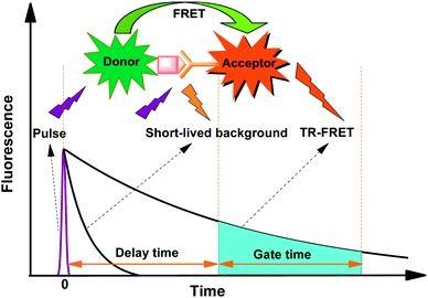
Many biological compounds present in cells, serum or other biological fluids are naturally fluorescent, and thus the use of conventional, prompt fluorophores leads to serious limitations in assay sensitivity due to the high background caused by the autofluorescence of the biological molecules to be assayed. The use of long-lived fluorophores combined with time-resolved detection (a delay between excitation and emission detection) minimizes prompt fluorescence interferences. Our trFluor™ Tb probes enable time-resolved fluorometry (TRF) for the assays that require high sensitivity. trFluor™ Tb probes have large Stokes shifts and extremely long emission half-lives when compared to more traditional fluorophores such as Alexa Fluor or cyanine dyes. Compared to the other TRF compounds, our trFluor™ Tb probes have relatively high stability, high emission yield and ability to be linked to biomolecules. This trFluor™ Tb goat anti-rabbit IgG (H+L) conjugate is commonly used as a second step reagent for indirect immunofluorescent staining, when used in conjunction with primary antibodies.

Spectrum


Open in Advanced Spectrum Viewer
spectrum

Spectral properties

Correction Factor (260 nm)0.942
Correction Factor (280 nm)0.797
Excitation (nm)333
Emission (nm)544

Product Family


NameExcitation (nm)Emission (nm)Extinction coefficient (cm -1 M -1)Correction Factor (260 nm)Correction Factor (280 nm)
trFluor™ Tb goat anti-mouse IgG (H+L)333544-0.9420.797
trFluor™ Tb goat anti-mouse IgG (H+L) *Cross Adsorbed*333544-0.9420.797
trFluor™ Eu goat anti-rabbit IgG (H+L)298617210000.9110.777
trFluor™ Eu goat anti-rabbit IgG (H+L) *Cross Adsorbed*298617210000.9110.777

Images


References


View all 61 references: Citation Explorer
Development of a time-resolved fluorescence resonance energy transfer assay for cyclin-dependent kinase 4 and identification of its ATP-noncompetitive inhibitors
Authors: Lo MC, Ngo R, Dai K, Li C, Liang L, Lee J, Emkey R, Eksterowicz J, Ventura M, Young SW, Xiao SH.
Journal: Anal Biochem (2012): 368
Time-Resolved Fluorescence Resonance Energy Transfer as a Versatile Tool in the Development of Homogeneous Cellular Kinase Assays
Authors: Saville L, Spais C, Mason JL, Albom MS, Murthy S, Meyer SL, Ator MA, Angeles TS, Husten J.
Journal: Assay Drug Dev Technol. (2012)
Oligomerization of the serotonin(1A) receptor in live cells: a time-resolved fluorescence anisotropy approach
Authors: Paila YD, Kombrabail M, Krishnamoorthy G, Chattopadhyay A.
Journal: J Phys Chem B (2011): 11439
A homogeneous single-label time-resolved fluorescence cAMP assay
Authors: Martikkala E, Rozw and owicz-Jansen A, Hanninen P, Petaja-Repo U, Harma H.
Journal: J Biomol Screen (2011): 356
Time-resolved fluorescence resonance energy transfer (TR-FRET) to analyze the disruption of EGFR/HER2 dimers: a new method to evaluate the efficiency of targeted therapy using monoclonal antibodies
Authors: Gaborit N, Larbouret C, Vallaghe J, Peyrusson F, Bascoul-Mollevi C, Crapez E, Azria D, Chardes T, Poul MA, Mathis G, Bazin H, Pelegrin A.
Journal: J Biol Chem (2011): 11337
Homogeneous time-resolved fluorescence-based assay to screen for ligands targeting the growth hormone secretagogue receptor type 1a
Authors: Leyris JP, Roux T, Trinquet E, Verdie P, Fehrentz JA, Oueslati N, Douzon S, Bourrier E, Lamarque L, Gagne D, Galleyr and JC, M'Kadmi C, Martinez J, Mary S, Baneres JL, Marie J.
Journal: Anal Biochem (2011): 253
A time-resolved fluorescence-resonance energy transfer assay for identifying inhibitors of hepatitis C virus core dimerization
Authors: Kota S, Scampavia L, Spicer T, Beeler AB, Takahashi V, Snyder JK, Porco JA, Hodder P, Strosberg AD.
Journal: Assay Drug Dev Technol (2010): 96
Time-resolved FRET fluorescence spectroscopy of visible fluorescent protein pairs
Authors: Visser AJ, Laptenok SP, Visser NV, van Hoek A, Birch DJ, Brochon JC, Borst JW.
Journal: Eur Biophys J (2010): 241
Steady-state and time-resolved fluorescence quenching with transition metal ions as short-distance probes for protein conformation
Authors: Posokhov YO, Kyrychenko A, Ladokhin AS.
Journal: Anal Biochem (2010): 284
Ligand regulation of the quaternary organization of cell surface M3 muscarinic acetylcholine receptors analyzed by fluorescence resonance energy transfer (FRET) imaging and homogeneous time-resolved FRET
Authors: Alvarez-Curto E, Ward RJ, Pediani JD, Milligan G.
Journal: J Biol Chem (2010): 23318