Products
Services
Resources
Selection Guides
About
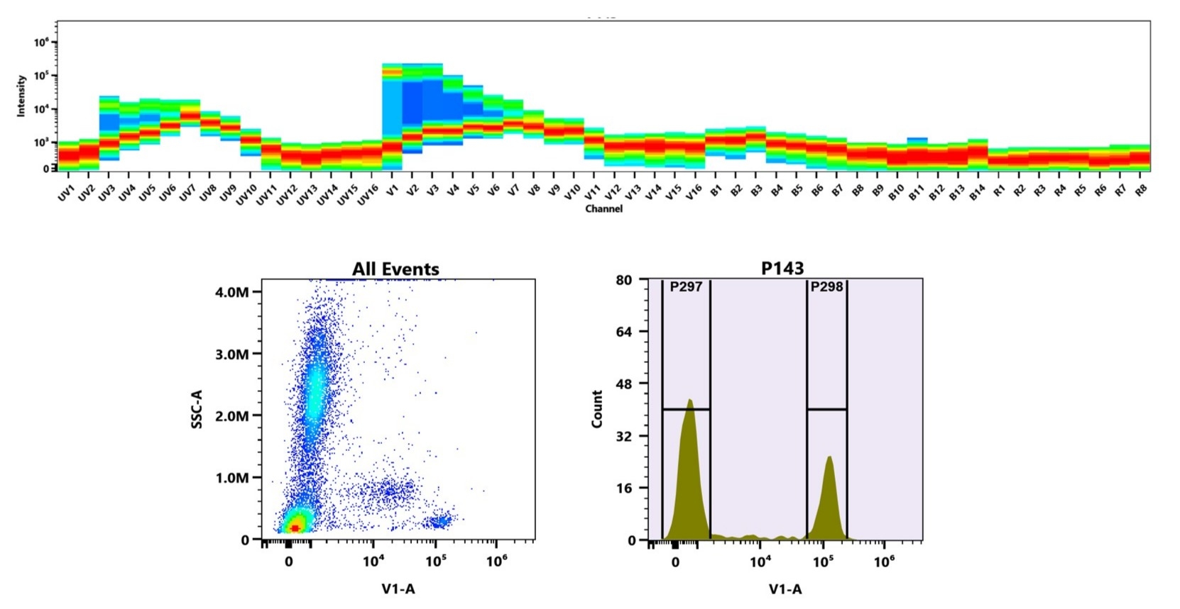
xtraFluor™ Violet 420 Maleimide
xtraFluor™ Violet 420 Maleimide enables thiol-selective antibody labeling with exceptional brightness for violet laser flow cytometry applications.
- Spectral Profile: Ex/Em ≈ 405/420 nm; optimized for the 405 nm violet laser
- Superior Dim Population Resolution: Brighter than Pacific Blue™ and BD Horizon™ V450, direct spectral match to BV421™
- Enhanced Stability Profile: Resists photobleaching and maintains fluorescence in standard buffers
- Maleimide Chemistry: Enables selective conjugation of antibodies
| Catalog | Size | Price | Quantity |
|---|---|---|---|
| 70802 | 100 ug | Price | |
| 70803 | 1 mg | Price |

Physical properties
| Molecular weight | 100000 |
| Solvent | DMSO |
Spectral properties
| Correction factor (280 nm) | 0.067 |
| Extinction coefficient (cm -1 M -1) | 3,000,000 |
| Excitation (nm) | 407 |
| Emission (nm) | 424 |
Usage and storage
| Intended use | Research Use Only (RUO) |
| Storage | Freeze (< -15 °C); Minimize light exposure |
Contact us
| Telephone | |
| Fax | |
| sales@aatbio.com | |
| International | See distributors |
| Bulk request | Inquire |
| Custom size | Inquire |
| Technical Support | Contact us |
| Request quotation | Request |
| Purchase order | Send to sales@aatbio.com |
| Shipping | Standard overnight for United States, inquire for international |
Page updated on July 15, 2026