Products
Services
Resources
Selection Guides
About
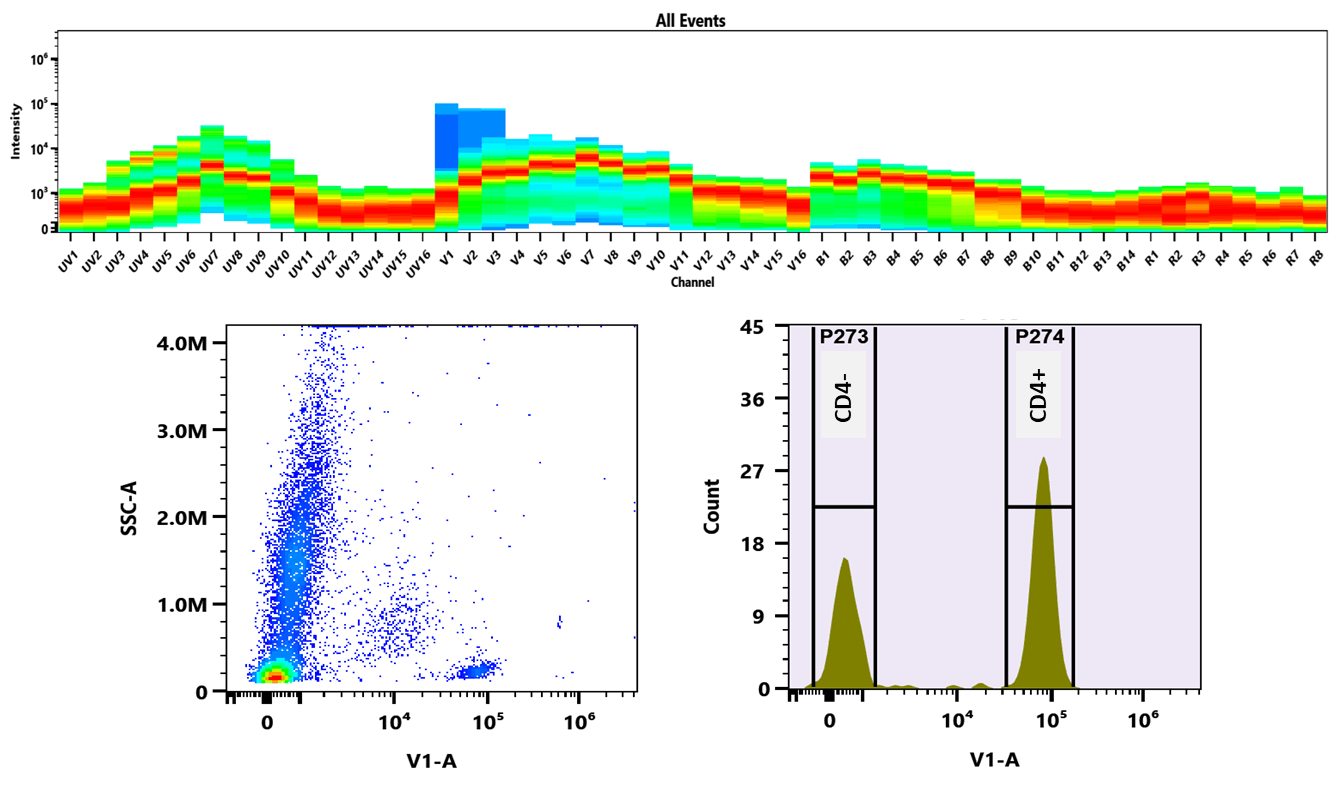
xtraFluor™ Violet 420 Labeling Dye
xtraFluor™ Violet 420 Labeling Dye is an exceptionally bright violet laser-excitable fluorophore for multicolor flow cytometry, outperforming Pacific Blue and BV421.
- Violet laser optimized: Excitation/emission maxima of ~405/420 nm matched for 405 nm violet laser workflows
- Bright violet fluorescence: Provides stronger fluorescence signal than Pacific Blue™ and BD Horizon™ V450, direct spectral match to BV421™
- Improved dim population resolution: Supports better discrimination of low-abundance or weakly stained cell populations
- Buccutite™ conjugation chemistry: Enables antibody labeling under mild conditions with high yields
- Enhanced workflow compatibility: Suitable for multicolor and spectral flow cytometry applications
| Catalog | Size | Price | Quantity |
|---|---|---|---|
| 70800 | 100 ug | Price | |
| 70801 | 1 mg | Price |

Spectral properties
| Correction factor (280 nm) | 0.067 |
| Extinction coefficient (cm -1 M -1) | 3,000,000 |
| Excitation (nm) | 407 |
| Emission (nm) | 424 |
Usage and storage
| Intended use | Research Use Only (RUO) |
Contact us
| Telephone | |
| Fax | |
| sales@aatbio.com | |
| International | See distributors |
| Bulk request | Inquire |
| Custom size | Inquire |
| Technical Support | Contact us |
| Request quotation | Request |
| Purchase order | Send to sales@aatbio.com |
| Shipping | Standard overnight for United States, inquire for international |
Page updated on July 15, 2026