Products
Services
Resources
Selection Guides
About
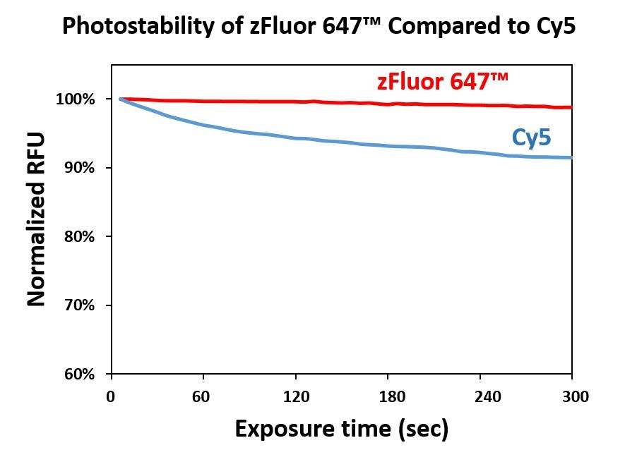
zFluor™ 647 succinimidyl ester
Our zFluor™ serial dyes are developed to have the possibly highest photostability at a given wavelength compared to the other similar wavelength dyes on the market. zFluor™ 647 has almost identical spectral properties to the popular Cy5® (GE Healthcare) and Alexa Fluor® 647 (ThermoFisher). Its high thermal and photostability makes it an excellent choice for single-molecule detection applications and high-resolution microscopy such as PALM, dSTORM and STED. Under the same test conditions, zFluor™ 647 has much higher ozone stability than Cy5® and Alexa Fluor® 647, making it a much better choice for microarray and other biochip-based applications. In addition, zFluor™-labeled oligonucleotides and peptides are much brighter and more photostable than the ones labeled by Alexa Fluor® 647 and Cy5®. This feature is extremely useful for fluorescence in-situ hybridization (FISH). In common with our iFluor® labels, the absorption and fluorescence of our zFluor™ 647 are independent of pH in the range of pH 2 to 11. It is well excited at 633 nm of He-Ne laser, the 647 nm line of the Krypton-Ion laser or a diode-laser emitting at 650 nm.
| Catalog | Size | Price | Quantity |
|---|---|---|---|
| 1500 | 1 mg | Price |

Physical properties
| Molecular weight | 853.04 |
| Solvent | DMSO |
Spectral properties
| Excitation (nm) | 648 |
| Emission (nm) | 668 |
Usage and storage
| Intended use | Research Use Only (RUO) |
| Storage | Freeze (< -15 °C); Minimize light exposure |
Contact us
| Telephone | |
| Fax | |
| sales@aatbio.com | |
| International | See distributors |
| Bulk request | Inquire |
| Custom size | Inquire |
| Technical Support | Contact us |
| Request quotation | Request |
| Purchase order | Send to sales@aatbio.com |
| Shipping | Standard overnight for United States, inquire for international |
Page updated on July 17, 2026