Products
Services
Resources
Selection Guides
About
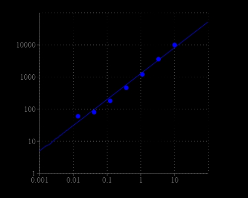
Amplite® Fluorimetric Hydrogen Peroxide Assay Kit
Red Fluorescence
Hydrogen peroxide (H2O2) is a reactive oxygen metabolic by-product that serves as a key regulator for a number of oxidative stress-related states. It is involved in a number of biological events that have been linked to asthma, atherosclerosis, diabetic vasculopathy, osteoporosis, a number of neurodegenerative diseases and Down's syndrome. Perhaps the most intriguing aspect of H2O2 biology is the recent report that antibodies have the capacity to convert molecular oxygen into hydrogen peroxide to contribute to the normal recognition and destruction processes of the immune system. Measurement of this reactive species will help to determine how oxidative stress modulates varied intracellular pathways. Amplite® Hydrogen Peroxide Assay Kit uses our Amplite® Red peroxidase substrate to quantify hydrogen peroxide in solutions and cell extracts. It can also be used to detect a variety of oxidase activities through enzyme-coupled reactions. The kit is an optimized 'mix and read' assay that is compatible with HTS liquid handling instruments.
| Catalog | Size | Price | Quantity |
|---|---|---|---|
| 11501 | 500 Tests | Price |

Spectral properties
| Excitation (nm) | 571 |
| Emission (nm) | 584 |
Storage, safety and handling
| H-phrase | H303, H313, H333 |
| Hazard symbol | XN |
| Intended use | Research Use Only (RUO) |
| R-phrase | R20, R21, R22 |
| Storage | Freeze (< -15 °C); Minimize light exposure |
| UNSPSC | 12171501 |
Instrument settings
| Fluorescence microplate reader | |
| Excitation | 540 nm |
| Emission | 590 nm |
| Cutoff | 570 nm |
| Recommended plate | Solid black |
Contact us
| Telephone | |
| Fax | |
| sales@aatbio.com | |
| International | See distributors |
| Bulk request | Inquire |
| Custom size | Inquire |
| Technical Support | Contact us |
| Request quotation | Request |
| Purchase order | Send to sales@aatbio.com |
| Shipping | Standard overnight for United States, inquire for international |
Page updated on June 21, 2026