logo
AAT Bioquest

Anti-D-Dimer antibody *Mouse anti-human, monoclonal IgG1*

Comparison between D-Dimer LFIA and diagnostic kit: 50 samples from donors were quantitatively tested using AAT Bioquest’s anti-D-Dimer antibodies in LFIA for the detection of D-dimer and a comparison kit. Results showed good correlation between both immunoassays, and the sensitivity reached to 0.2μg/ml.
Calibration curve for D-Dimer: human D-Dimer was reacted with anti-human D-Dimer antibody-coated latex, resulting in agglutination and increase in turbidity. Changes in turbidity were monitored with a spectrometer to quantitatively measure the D-Dimer concentration in the sample. The linear range is about 30μg/ml.
Comparison between D-Dimer LETIA and diagnostic kit: samples from donors(n=51) were detected using AAT Bioquest’s anti-D-Dimer antibodies in LETIA and a diagnostic kit. Results showed good correlation between both assays, and the sensitivity reached to 0.2μg/ml.
Ordering information
Price
Catalog Number
Unit Size
Quantity
Add to cart
Additional ordering information
Telephone1-800-990-8053
Fax1-800-609-2943
Emailsales@aatbio.com
InternationalSee distributors
Bulk requestInquire
Custom sizeInquire
ShippingStandard overnight for United States, inquire for international
Request quotation
Physical properties
SolventWater
Storage, safety and handling
Intended useResearch Use Only (RUO)
StorageFreeze (< -15 °C)

OverviewpdfSDSpdfProtocol


Other names
Host
mouse
Reactivity
human
Application
ELISA; LFIA; LETIA
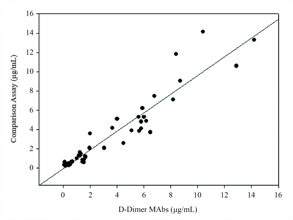
D-dimer is a small protein fragment, or fibrin degradation product (FDP), present in blood after a blood clot has been degraded by fibrinolysis. It is composed of two D fragments of the fibrin protein joined together by a cross-link. Elevated d-dimer levels are a key indicator or thrombotic events, indicating excess fibrinolysis, following activation of coagulation. In clinical biology, it is used in the diagnosis of the blood disorder disseminated intravascular coagulation. Anti-D-Dimer monoclonal antibodies are highly sensitive for the detection of human D-dimer immunogens. They can be used in a broad range of immunoassays such as lateral-flow immunoassays (LFIA) and in IVD assay development.

Conjugation


We provide custom conjugation services for this antibody (eg. labeling of Anti-D-Dimer antibody *Mouse anti-human, monoclonal IgG1* with HRP). A list of available labels can be found in the table below:

AFAF350, AF488, AF555, AF594, AF647, AF680, AF700, AF750
ProteinsHRP, Alkaline Phosphatase, Streptavidin
TandemsAPC, APC/Cy7, APC/AF750, APC/iFluor™ 700, APC/iFluor™ 750, PE, PE/Cy5, PE/Cy7, PE/AF610, PE/AF700, PE/iFluor™ 594, PE/iFluor™ 647, PE/iFluor™ 700, PE/iFluor™ 750, PE/Texas Red®, PerCP, PerCP/Cy5.5
Small MoleculesBiotin
Traditional DyesFITC (fluorescein), TRITC, PacBlue, PacOrange, Cy3, Cy5
iFluor350, 405, 430, 450, 488, 514, 532, 546, 555, 560, 568, 594, 610, 633, 647, 660, 670, 680, 700, 710, 750, 790, 800, 810, 820, 840, 860, A7
mFluorUV375, UV460, Violet 450, Violet 500, Violet 510, Violet 540, Blue 570, Green 620, Red 700, Red 780

For additional information about custom conjugations, please consult our services page here.

Images