Products
Services
Resources
Selection Guides
About
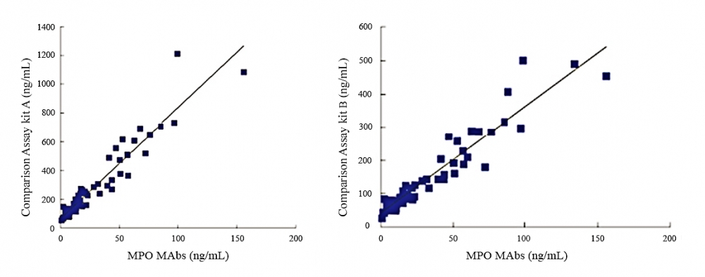
Anti-Myeloperoxidase (MPO) antibody
Mouse anti-human, monoclonal IgG2b
Myeloperoxidase (MPO) is a 150 kDa cationic hemoprotein that consists of two covalently bound subunits each containing a heavy chain (60 kDa) and a light chain (15 kDa). MPO is abundantly expressed in neutrophils and is released during degranulation. It plays a vital role in inflammatory responses by catalyzing chloride ion oxidation to produce hypochlorous acid which is used by neutrophils to kill bacteria and other pathogens. MPO is also known to cause oxidative modification of low density lipoproteins to a high uptake form. This is considered to be a key event in the promotion of atherogenesis and is linked to the progression of cardiovascular diseases. As such, MPO is considered to be a promising cardiovascular biomarker as elevated MPO levels indicates a high risk for atherosclerosis and coronary heart disease (CHD). Anti-MPO monoclonal antibodies are highly sensitive for the detection of human MPO. They can be used for a broad range of immunoassays such as latex enhanced turbidimetric immunoassays (LETIA), chemiluminescent immunoassays (CLIA) and ELISA.
| Catalog | Size | Price | Quantity |
|---|---|---|---|
| V100095 | 50 ug | Price | |
| V100096 | 1 mg | Price |
Physical properties
| Solvent | Water |
Antibody properties
| Host | Mouse |
| Reactivity | Human |
| Application | LETIA, ELISA, CLIA, LFIA |
Storage, safety and handling
| Intended use | Research Use Only (RUO) |
| Storage | Freeze (< -15 °C) |
Contact us
| Telephone | |
| Fax | |
| sales@aatbio.com | |
| International | See distributors |
| Bulk request | Inquire |
| Custom size | Inquire |
| Technical Support | Contact us |
| Request quotation | Request |
| Purchase order | Send to sales@aatbio.com |
| Shipping | Standard overnight for United States, inquire for international |
Page updated on July 10, 2026