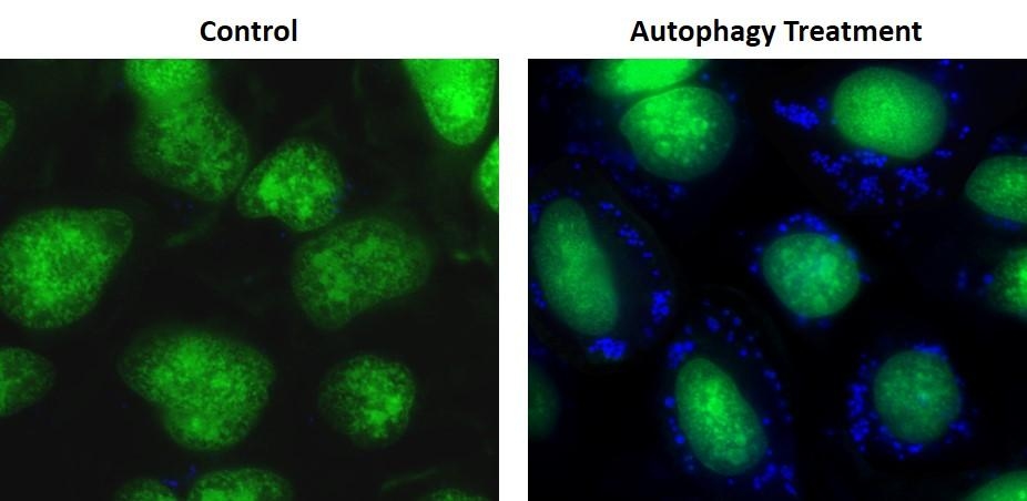
Cell Meter™ Autophagy Fluorescence Imaging Kit
Ordering information
Price | |
Catalog Number | |
Unit Size | |
Quantity |
Additional ordering information
Telephone | 1-800-990-8053 |
Fax | 1-800-609-2943 |
sales@aatbio.com | |
International | See distributors |
Bulk request | Inquire |
Custom size | Inquire |
Shipping | Standard overnight for United States, inquire for international |
Storage, safety and handling
H-phrase | H303, H313, H333 |
Hazard symbol | XN |
Intended use | Research Use Only (RUO) |
R-phrase | R20, R21, R22 |
UNSPSC | 12352200 |
Related products
Overview | ![]() ![]() |
Autophagy is one of the major pathways for degradation of intracellular macromolecules in animal cells. The process of autophagy involves the sequestration of cytoplasmic materials and intracellular organelles in a membrane-bounded vacuole called autophagosome, the fusion of the autophagosome with lysosomes, and the subsequent degradation of sequestered materials. Cell Meter™ autophagy fluorescence imaging kit employs Autophagy Super Blue™ as a specific autophagosome marker to analyze the activity of autophagy. The assay is optimized for direct detection of autophagy in both detached and attached cells. The kit provides all the essential components for the assay protocol. The Cell Meter™ autophagy fluorescence imaging kit is optimized for fluorescence microscope. It gives much higher selectivity than other commercially available autophagy probes.
Platform
Fluorescence microscope
Excitation | DAPI filter |
Emission | DAPI filter |
Recommended plate | Black wall/clear bottom |
Components
Example protocol
AT A GLANCE
Protocol summary
- Prepare cells with your test compounds at the density of 1 - 2 × 10⁴ cells/well
- Add Autophagy Super Blue™ working solution
- Incubate at 37°C for 15 - 60 minutes
- Wash cells with Wash Buffer
- Monitor the fluorescence increase at Ex/Em= 330/520 nm (Cutoff = 475 nm), fluorescence microscope with DAPI filter set
Important notes
Thaw all the components at room temperature before starting the experiment.
PREPARATION OF WORKING SOLUTION
Add 20 μL of 500X Autophagy Super Blue™ (Component A) to 10 mL of Stain Buffer (Component B) and mix well to make Autophagy Super Blue™ working solution. Protect from light. Note: 20 μL of 500X Autophagy Super Blue™ (Component A) is enough for one 96-well plate.
SAMPLE EXPERIMENTAL PROTOCOL
- Culture cells to a density optimal for autophagy induction according to your specific induction protocol (about 1 - 2 × 10⁴ cells/ well/96-well plate). At the same time, culture a non-induced negative control cell population at the same density as the induced population for every labeling condition.
- Remove medium.
- Add 100 µL/well (96-well plate) or 25 µL/well (384-well plate) of Autophagy Super Blue™ working solution into each well.
- Incubate the cells in a 37°C, 5% CO2 incubator for 15 to 60 minutes. Note: The appropriate incubation time depends on the individual cell type and cell concentration used. Optimize the incubation time for each experiment.
- Wash the cells with Wash Buffer (Component C) for 3 - 4 times, then add 100 µL Wash Buffer (Component C) to each well. Note: It is recommended to increase either the labeling concentration or the incubation time to allow the dye to accumulate if the cells do not appear to be sufficiently stained.
- Monitor the fluorescence intensity with a fluorescence microplate reader at Ex/Em = 330/520 nm (Cutoff = 475 nm), a fluorescence microscope with DAPI filter set.
Images

Figure 1. Autophagy Super Blue™ labeled vesicles were induced by starvation in HeLa cells. HeLa cells were incubated in a regular DMEM medium (Left: Control) or in 1X HBSS buffer with 5% serum (Right: Autophagy Treatment) for 16 hours. Both control and treated cells were incubated with Autophagy Super Blue™ working solution for 20 minutes in a 37 °C, 5% CO2 incubator, and washed 3 times with wash buffer. Cells were imaged immediately under a fluorescence microscope with a DAPI channel (blue). Cell nuclei were stained with Nuclear Green™ LCS1 (green).
Citations
View all 7 citations: Citation Explorer
Immune privilege of adipocyte mitochondria protects from obesity
Authors: Hoang, Anh Cuong and Yu, Haidong and Lin, Ya-Tin and Chen, Jin-Chung and Chen, Chia-Chun and Diedrich, Victoria and Herwig, Annika and Landgraf, Kathrin and K{\"o}rner, Antje and Roszer, Tam{\'a}s
Journal: (2021)
Authors: Hoang, Anh Cuong and Yu, Haidong and Lin, Ya-Tin and Chen, Jin-Chung and Chen, Chia-Chun and Diedrich, Victoria and Herwig, Annika and Landgraf, Kathrin and K{\"o}rner, Antje and Roszer, Tam{\'a}s
Journal: (2021)
TCP1 increases drug resistance in acute myeloid leukemia by suppressing autophagy via activating AKT/mTOR signaling
Authors: Chen, Xiaofang and Chen, Xianling and Huang, Yiping and Lin, Jia and Wu, Yong and Chen, Yuanzhong
Journal: Cell death \& disease (2021): 1--11
Authors: Chen, Xiaofang and Chen, Xianling and Huang, Yiping and Lin, Jia and Wu, Yong and Chen, Yuanzhong
Journal: Cell death \& disease (2021): 1--11
Primary cilia and autophagy interaction is involved in mechanical stress mediated cartilage development via ERK/mTOR axis
Authors: Xiang, Wei and Jiang, Ting and Hao, Xiaoxia and Wang, Rui and Yao, Xudong and Sun, Kai and Guo, Fengjing and Xu, Tao
Journal: Life Sciences (2019)
Authors: Xiang, Wei and Jiang, Ting and Hao, Xiaoxia and Wang, Rui and Yao, Xudong and Sun, Kai and Guo, Fengjing and Xu, Tao
Journal: Life Sciences (2019)
Tanshinone IIA ameliorates oxaliplatin-induced neurotoxicity via mitochondrial protection and autophagy promotion
Authors: Cheng, Weiting and Xiang, Wei and Wang, Shan and Xu, Kai
Journal: Am J Transl Res (2019): 3140--3149
Authors: Cheng, Weiting and Xiang, Wei and Wang, Shan and Xu, Kai
Journal: Am J Transl Res (2019): 3140--3149
Methods for Measuring Autophagy Levels in Disease
Authors: Phadwal, Kanchan and Kurian, Dominic
Journal: (2017): 195--211
Authors: Phadwal, Kanchan and Kurian, Dominic
Journal: (2017): 195--211
High glucose induces bone marrow-derived mesenchymal stem cell senescence by upregulating autophagy
Authors: Chang, Tzu-Ching and Hsu, Min-Fen and Wu, Kenneth K
Journal: PloS one (2015): e0126537
Authors: Chang, Tzu-Ching and Hsu, Min-Fen and Wu, Kenneth K
Journal: PloS one (2015): e0126537
Licochalcone A induces autophagy through PI3K/Akt/mTOR inactivation and autophagy suppression enhances Licochalcone A-induced apoptosis of human cervical cancer cells
Authors: Tsai, Jen-Pi and Lee, Chien-Hsing and Ying, Tsung-Ho and Lin, Chu-Liang and Lin, Chia-Liang and Hsueh, Jung-Tsung and Hsieh, Yi-Hsien
Journal: Oncotarget (2015): 28851
Authors: Tsai, Jen-Pi and Lee, Chien-Hsing and Ying, Tsung-Ho and Lin, Chu-Liang and Lin, Chia-Liang and Hsueh, Jung-Tsung and Hsieh, Yi-Hsien
Journal: Oncotarget (2015): 28851
References
View all 28 references: Citation Explorer
beta-Elemene induces apoptosis as well as protective autophagy in human non-small-cell lung cancer A549 cells
Authors: Liu J, Hu XJ, Jin B, Qu XJ, Hou KZ, Liu YP.
Journal: J Pharm Pharmacol (2012): 146
Authors: Liu J, Hu XJ, Jin B, Qu XJ, Hou KZ, Liu YP.
Journal: J Pharm Pharmacol (2012): 146
Tgf-beta1 induces autophagy and promotes apoptosis in renal tubular epithelial cells
Authors: Xu Y, Yang S, Huang J, Ruan S, Zheng Z, Lin J.
Journal: Int J Mol Med. (2012)
Authors: Xu Y, Yang S, Huang J, Ruan S, Zheng Z, Lin J.
Journal: Int J Mol Med. (2012)
High-Throughput Screening for AntiInfluenza A Virus Drugs and Study of the Mechanism of Procyanidin on Influenza A VirusInduced Autophagy
Authors: Dai J, Wang G, Li W, Zhang L, Yang J, Zhao X, Chen X, Xu Y, Li K.
Journal: J Biomol Screen. (2012)
Authors: Dai J, Wang G, Li W, Zhang L, Yang J, Zhao X, Chen X, Xu Y, Li K.
Journal: J Biomol Screen. (2012)
An autophagy inhibitor enhances the inhibition of cell proliferation
Authors: Yao F, Wang G, Wei W, Tu Y, Tong H, Sun S.
Journal: Mol Med Report (2012): 84
Authors: Yao F, Wang G, Wei W, Tu Y, Tong H, Sun S.
Journal: Mol Med Report (2012): 84
Inhibition of induced autophagy increases apoptosis of Nara-H cells
Authors: Nakamura O, Hitora T, Akisue T, Kawamoto T, Yamagami Y, Yamamoto T.
Journal: Int J Oncol (2011): 1545
Authors: Nakamura O, Hitora T, Akisue T, Kawamoto T, Yamagami Y, Yamamoto T.
Journal: Int J Oncol (2011): 1545
A new fluorescence-based assay for autophagy
Authors: Proikas-Cezanne T, Codogno P.
Journal: Chem Biol (2011): 940
Authors: Proikas-Cezanne T, Codogno P.
Journal: Chem Biol (2011): 940
Reactive oxygen species contribute to oridonin-induced apoptosis and autophagy in human cervical carcinoma HeLa cells
Authors: Zhang YH, Wu YL, Tashiro S, Onodera S, Ikejima T.
Journal: Acta Pharmacol Sin (2011): 1266
Authors: Zhang YH, Wu YL, Tashiro S, Onodera S, Ikejima T.
Journal: Acta Pharmacol Sin (2011): 1266
Salvianolic acid B inhibits autophagy and protects starving cardiac myocytes
Authors: Han X, Liu JX, Li XZ.
Journal: Acta Pharmacol Sin (2011): 38
Authors: Han X, Liu JX, Li XZ.
Journal: Acta Pharmacol Sin (2011): 38
Quantification of nonselective bulk autophagy in S. cerevisiae using Pgk1-GFP
Authors: Welter E, Thumm M, Krick R.
Journal: Autophagy (2010): 794
Authors: Welter E, Thumm M, Krick R.
Journal: Autophagy (2010): 794
Concomitant inhibition of AKT and autophagy is required for efficient cisplatin-induced apoptosis of metastatic skin carcinoma
Authors: Claerhout S, Verschooten L, Van Kelst S, De Vos R, Proby C, Agostinis P, Garmyn M.
Journal: Int J Cancer (2010): 2790
Authors: Claerhout S, Verschooten L, Van Kelst S, De Vos R, Proby C, Agostinis P, Garmyn M.
Journal: Int J Cancer (2010): 2790
Application notes
FAQ
Can I use any of your Cell Meter™ Autophagy Assay Kits with a flow cytometer?
How does the autophagy marker in your Cell Meter™ Autophagy Assay Kit (Cat#23000) work?
How do cells protect themselves against cellular stress?
What are the most common types of cell death?
Are Cell Navigator® Cell Plasma Membrane Staining Kits suitable for cell culture medium samples?
How does the autophagy marker in your Cell Meter™ Autophagy Assay Kit (Cat#23000) work?
How do cells protect themselves against cellular stress?
What are the most common types of cell death?
Are Cell Navigator® Cell Plasma Membrane Staining Kits suitable for cell culture medium samples?