Cell Meter™ Fluorimetric Cellular Lipid Peroxidation Assay Kit
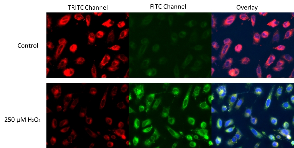
Lipid peroxidation is the oxidative degradation of cellular lipid by reactive oxygen species (ROS). This process can lead to not only disruption of the cellular membrane integrity, but also inactivation of membrane-bound receptors. It is one of the main causes of free radical-mediated damages in cells. Cell Meter™ Fluorimetric Cellular Lipid Peroxidation Assay Kit provides a sensitive method for monitoring lipid peroxidation. The kit uses our sensitive ratiometric lipid peroxidation sensor, Lipoxite™ R590/G520 that changes its red fluorescence from red to green upon peroxidation by ROS in cells, this peroxidation-dependent shift enables the ratiometric measurement of lipid peroxidation. Our kit includes H2O2 as a positive control treatment to induce lipid peroxidation.
![HeLa cells were stained with 1X Lipoxite™ R590/G525 for 30 mins in complete growth medium at 37°C. For H<sub>2</sub>O<sub>2</sub> treatment, approximately 250 µM of H<sub>2</sub>O<sub>2</sub> were added to the cells and incubated for 30 mins. The cells were then incubated with 1X Lipoxite™ R590/G525, and stained with Hoechst 33342 during the last 10 mins of incubation. The cells were washed 3 times with HHBS and imaged with a Keyence fluorescent microscope. With H<sub>2</sub>O<sub>2</sub> treatment, a clear shift of fluorescence signal of red to green was observed.](/_next/image?url=https%3A%2F%2Fimages.aatbio.com%2Fproducts%2Ffigures-and-data%2Fcell-meter-fluorimetric-cellular-lipid-peroxidation-assay-kit%2Ffigure-for-cell-meter-fluorimetric-cellular-lipid-peroxidation-assay-kit_8GDMm.jpg&w=640&q=75)
![HeLa cells were stained with 1X Lipoxite™ R590/G525 for 30 mins in complete growth medium at 37°C. For H<sub>2</sub>O<sub>2</sub> treatment, approximately 250 µM of H<sub>2</sub>O<sub>2</sub> were added to the cells and incubated for 30 mins. The cells were then incubated with 1X Lipoxite™ R590/G525, and stained with Hoechst 33342 during the last 10 mins of incubation. The cells were washed 3 times with HHBS and imaged with a Keyence fluorescent microscope. With H<sub>2</sub>O<sub>2</sub> treatment, a clear shift of fluorescence signal of red to green was observed.](/_next/image?url=https%3A%2F%2Fimages.aatbio.com%2Fproducts%2Ffigures-and-data%2Fcell-meter-fluorimetric-cellular-lipid-peroxidation-assay-kit%2Ffigure-for-cell-meter-fluorimetric-cellular-lipid-peroxidation-assay-kit_8GDMm.jpg&w=128&q=25)
![Jurkat cells were stained with 1X Lipoxite™ R590/G525 for 30 mins in complete growth medium at 37°C. For H<sub>2</sub>O<sub>2</sub> treatment, approximately 250 µM of H<sub>2</sub>O<sub>2</sub> were added to the cells and incubated for 30 mins. The cells were then incubated with 1X Lipoxite™ R590/G525, and analyzed with a flow cytometer through FITC (488/530 nm) and PE (488/572 nm) channels. The data are represented as the ratios of red (PE)/green (FITC) fluorescence intensities. The ratio of red/green decreases in H<sub>2</sub>O<sub>2</sub> treated cells indicating the presence of H<sub>2</sub>O<sub>2</sub> induced lipid peroxidation.](/_next/image?url=https%3A%2F%2Fimages.aatbio.com%2Fproducts%2Ffigures-and-data%2Fcell-meter-fluorimetric-cellular-lipid-peroxidation-assay-kit%2Ffigure-for-cell-meter-fluorimetric-cellular-lipid-peroxidation-assay-kit_fypeM.jpg&w=128&q=25)
Example protocol
AT A GLANCE
Protocol summary
- Plate cells at desired confluency.
- Treat cells with compound of interest and incubate.
- Add Lipoxite™ R590/G525.
- Remove media, and wash with PBS.
- Analyze sample by fluorescence microscope or flow cytometer through FITC/TRITC or FITC/PE channels.
Important
Thaw one of each kit component at room temperature before starting the experiment.
PREPARATION OF WORKING SOLUTION
Prepare 10X working solution of Lipoxite™ R590/G525 by making 1:50 dilution of 500X Lipoxite™ R590/G525 (Component A) into HHBS (Component B)
For guidelines on cell sample preparation, please visit
https://www.aatbio.com/resources/guides/cell-sample-preparation.html
SAMPLE EXPERIMENTAL PROTOCOL
- Grow cells at desired density and incubate overnight in a humidified chamber at 37°C with 5% CO2.
- Treat cells with test compounds as desired. Note: For a positive control, add hydrogen peroxide (Component C) to the cells at a final concentration of ~250 µM (1X) for 30 minutes.
- Add 10X Lipoxite™ R590/G525 to the cells at a final concentration of 1X (for example add 10 uL to 90 uL of the cells).
- Incubate the cells for 30 min at 37°C with 5% CO2 cell incubator.
- Remove media and wash cells with HHBS (Component B) or DPBS for three times.
- Monitor fluorescence of cells with a fluorescence microscope or flow cytometer through FITC/TRITC or FITC/PE channels within 2 hours of staining.
Citations
View all 19 citations: Citation Explorer
PDT-Enhanced Ferroptosis by a Polymer Nanoparticle with pH-Activated Singlet Oxygen Generation and Superb Biocompatibility for Cancer Therapy
Authors: Li, Jing and Li, Junhua and Pu, Yuji and Li, Sai and Gao, Wenxia and He, Bin
Journal: Biomacromolecules (2021): 1167--1176
Authors: Li, Jing and Li, Junhua and Pu, Yuji and Li, Sai and Gao, Wenxia and He, Bin
Journal: Biomacromolecules (2021): 1167--1176
Using fluorescence-activated flow cytometry to determine reactive oxygen species formation and membrane lipid peroxidation in viable boar spermatozoa
Authors: Guthrie, H. D., Welch, G. R.
Journal: Methods Mol Biol (2010): 163-71
Authors: Guthrie, H. D., Welch, G. R.
Journal: Methods Mol Biol (2010): 163-71
Use of fluorescence-activated flow cytometry to determine membrane lipid peroxidation during hypothermic liquid storage and freeze-thawing of viable boar sperm loaded with 4, 4-difluoro-5-(4-phenyl-1,3-butadienyl)-4-bora-3a,4a-diaza-s-indacene-3-undecanoic
Authors: Guthrie, H. D., Welch, G. R.
Journal: J Anim Sci (2007): 1402-11
Authors: Guthrie, H. D., Welch, G. R.
Journal: J Anim Sci (2007): 1402-11
Fluorescence microplate reader measurement of tissue susceptibility to lipid peroxidation
Authors: Zhang, J. G., Fariss, M. W.
Journal: Curr Protoc Toxicol (2004): Unit17 3
Authors: Zhang, J. G., Fariss, M. W.
Journal: Curr Protoc Toxicol (2004): Unit17 3
Response to ozone in two lettuce varieties on chlorophyll a fluorescence, photosynthetic pigments and lipid peroxidation
Authors: Calatayud, A., Barreno, E.
Journal: Plant Physiol Biochem (2004): 549-55
Authors: Calatayud, A., Barreno, E.
Journal: Plant Physiol Biochem (2004): 549-55