DAPI
4,6-Diamidino-2-phenylindole, dihydrochloride; CAS 28718-90-3
DAPI is a blue fluorescent dye that stains cell nuclei in fluorescence microscopy by binding to A-T rich regions of dsDNA.
Excitation and emission
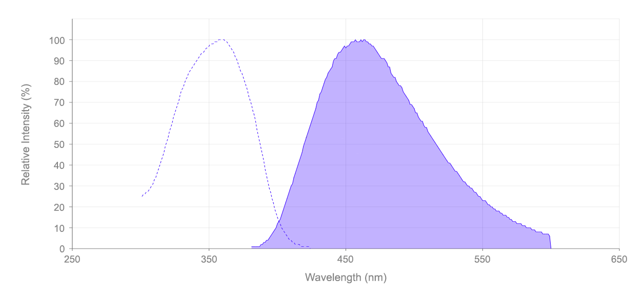
DAPI, or 4',6-diamidino-2-phenylindole, is a fluorescent compound with an excitation peak at 359 nm and an emission peak at 457 nm. It is well excited by the 405 nm violet laser or the 355 nm UV laser. The emission can be captured by a 450/50 nm bandpass filter or using the DAPI filter set configuration on fluorescence microscopes.Staining principle and mechanism
DAPI is an intercalating dye, binding to the minor groove of A-T rich regions in double stranded DNA (dsDNA). This preference possibly stems from the more negative electrostatic potential of these areas, which would facilitate the formation of hydrogen bonds and hydrophobic interactions between the dye molecules and DNA base pairs. Upon binding to these regions, the quantum yield of DAPI increases more than 20-fold, resulting in bright blue fluorescence when excited by a violet (405 nm) laser. Intercalation with G-C regions, on the other hand, results in minimal to no fluorescence, while binding to A-U rich regions of RNA produces fluorescence but at a much weaker intensity, with the quantum yield of DAPI increasing only about 5-fold.Permeability
DAPI is cell impermeable at low concentrations. Therefore, it is often used to stain dead, necrotic or fixed cells, where membrane integrity is compromised. At higher concentrations, it is semi-permeable, with the ability to stain live cells. However, due to its high cytotoxicity, the staining of live cells with DAPI is not recommended. Because DAPI primarily stains cell nuclei, where the concentration of dsDNA is highest, it is often used as a co-stain or counterstain for applications such as cell imaging, cell counting and cell sorting, and it has been used to stain a variety of cell types, such as animal, bacterial and fungal.Protocol and usage
Typical DAPI staining can be performed in 5 to 15 minutes, depending on the protocol used. The staining may be stable for several hours to days under appropriate conditions, though fluorescence intensity may decrease over time. Samples stained with DAPI should be washed after staining to remove any unbound dye and to reduce background fluorescence. To fix cells after staining, paraformaldehyde or other fixative solutions may be used. The staining of cells with DAPI is irreversible.DAPI vs Hoechst
Some alternatives to DAPI include the Hoechst family of dyes. While both bind to DNA, Hoechst stains tend to have lower cytotoxicity and greater membrane permeability, allowing for their use in staining live cells. DAPI, having poor membrane permeability and greater toxicity, is more often used to stain dead or fixed cells.
Kapuscinski, J. (1995). DAPI: a DNA-specific fluorescent probe. Biotechnic & histochemistry, 70(5), 220-233.
DAPI working concentration
DAPI has a molecular weight of 350.25 daltons. It has moderate solubility in water (20 mg / mL) and yields a clear yellow solution when dissolved. To make a working solution of DAPI, first add deionized water (diH2O) or dimethylformamide (DMF) to the powdered form. Sonication may be necessary for contents to fully dissolve. After, dilute DAPI solution with PBS to required concentrations. Typical staining protocols use concentrations between 0.5 µM to 5 µM. Once prepared, the solution is stable for several weeks when stored in a dark location at 2 to 6 °C. For longer storage times, keep at < -15 °C.
DAPI flow cytometry protocol
-
Prepare staining buffer sufficient for samples (1 mL per cell sample). Staining buffer is prepared using the following recipe:
100 mM Tris, pH 7.4
150 mM NaCl
1 mM CaCl2
0.5 mM MgCl2
0.1% Nonidet P-40 - Use the fixation protocol appropriate for your sample.
- Dilute DAPI solution with prepared staining buffer to 3 µM.
- Centrifuge cell suspension and discard the supernatant. Tap to loosen the pellet and add 1 mL of DAPI staining solution.
- Incubate at room temperature for 15 minutes.
- Analyze using flow cytometry with 405 nm excitation laser and 450/50 nm bandpass filter (or equivalent channel).
General staining protocol
- Use the fixation protocol appropriate for your sample.
- Pellet cells by centrifugation and resuspend cells in buffered salt solutions or media, with optimal dye binding at pH 7.4
- Add DAPI stain using concentrations between 0.5 µM to 5 µM.
- Incubate for 15 to 60 minutes.
- Image the cells using a fluorescence microscope with the DAPI filter set.
Calculators
Common stock solution preparation
Table 1. Volume of Water needed to reconstitute specific mass of DAPI [4,6-Diamidino-2-phenylindole, dihydrochloride] *CAS 28718-90-3* to given concentration. Note that volume is only for preparing stock solution. Refer to sample experimental protocol for appropriate experimental/physiological buffers.
0.1 mg | 0.5 mg | 1 mg | 5 mg | 10 mg | |
1 mM | 285.51 µL | 1.428 mL | 2.855 mL | 14.276 mL | 28.551 mL |
5 mM | 57.102 µL | 285.51 µL | 571.021 µL | 2.855 mL | 5.71 mL |
10 mM | 28.551 µL | 142.755 µL | 285.51 µL | 1.428 mL | 2.855 mL |
Molarity calculator
Enter any two values (mass, volume, concentration) to calculate the third.
Mass (Calculate) | Molecular weight | Volume (Calculate) | Concentration (Calculate) | Moles | ||||
/ | = | x | = |
Spectrum
Open in Advanced Spectrum Viewer
Citations
View all 87 citations: Citation Explorer
An NIR-responsive hydrogel loaded with polydeoxyribonucleotide nano-vectors for enhanced chronic wound healing
Authors: Sun, Yanzhen and Li, Yao and Ding, Xiaokang and Xu, Pan and Jing, Xiaodong and Cong, Hailin and Hu, Hao and Yu, Bing and Xu, Fu-Jian
Journal: Biomaterials (2024): 122789
Authors: Sun, Yanzhen and Li, Yao and Ding, Xiaokang and Xu, Pan and Jing, Xiaodong and Cong, Hailin and Hu, Hao and Yu, Bing and Xu, Fu-Jian
Journal: Biomaterials (2024): 122789
Emergence of large-scale cell death through ferroptotic trigger waves
Authors: Co, Hannah KC and Wu, Chia-Chou and Lee, Yi-Chen and Chen, Sheng-hong
Journal: Nature (2024): 1--9
Authors: Co, Hannah KC and Wu, Chia-Chou and Lee, Yi-Chen and Chen, Sheng-hong
Journal: Nature (2024): 1--9
Deciphering the Role of Ferroptosis in Rheumatoid Arthritis: Synovial Transcriptome Analysis and Immune Infiltration Correlation
Authors: Wang, Hongli and Zhang, Miaomiao and Hu, Yiping and He, Juan and Zhong, Yuchao and Dai, Yong and Wang, Qingwen
Journal: Heliyon (2024)
Authors: Wang, Hongli and Zhang, Miaomiao and Hu, Yiping and He, Juan and Zhong, Yuchao and Dai, Yong and Wang, Qingwen
Journal: Heliyon (2024)
The mesenchymal stromal cell (MSC) secretome promotes tissue regeneration and increases macrophage infiltration in acute and MRSA-infected skin wounds in vivo
Authors: Rajesh, Aarthi and Ju, Esther Da Eun and Oxford, Kelly A and Harman, Rebecca M and Van de Walle, Gerlinde R
Journal: Cytotherapy (2024)
Authors: Rajesh, Aarthi and Ju, Esther Da Eun and Oxford, Kelly A and Harman, Rebecca M and Van de Walle, Gerlinde R
Journal: Cytotherapy (2024)
Adipose Tissue and Translation Machinery in Metabolic Regulation
Authors: De Siqueira, Mirian Krystel
Journal: (2024)
Authors: De Siqueira, Mirian Krystel
Journal: (2024)
References
View all 134 references: Citation Explorer
Reliable DNA ploidy determination in dehydrated tissues of vascular plants by DAPI flow cytometry--new prospects for plant research
Authors: Suda J, Travnicek P.
Journal: Cytometry A (2006): 273
Authors: Suda J, Travnicek P.
Journal: Cytometry A (2006): 273
Identification of all pachytene bivalents in the common shrew using DAPI-staining of synaptonemal complex spreads
Authors: Belonogova NM, Karamysheva TV, Biltueva LS, Perepelov EA, Minina JM, Polyakov AV, Zhdanova NS, Rubtsov NB, Searle JB, Borodin PM.
Journal: Chromosome Res (2006): 673
Authors: Belonogova NM, Karamysheva TV, Biltueva LS, Perepelov EA, Minina JM, Polyakov AV, Zhdanova NS, Rubtsov NB, Searle JB, Borodin PM.
Journal: Chromosome Res (2006): 673
Physical localization of ribosomal genes and chromosome DAPI banding by in situ hybridization in Medicago sativa L
Authors: Chen JM, Hong YH, Wang YP, Bowley S, Wan JM.
Journal: Yi Chuan (2006): 184
Authors: Chen JM, Hong YH, Wang YP, Bowley S, Wan JM.
Journal: Yi Chuan (2006): 184
DNA staining with the fluorochromes EtBr, DAPI and YOYO-1 in the comet assay with tobacco plants after treatment with ethyl methanesulphonate, hyperthermia and DNase-I
Authors: Gichner T, Mukherjee A, Veleminsky J.
Journal: Mutat Res (2006): 17
Authors: Gichner T, Mukherjee A, Veleminsky J.
Journal: Mutat Res (2006): 17
Investigation of chromosomes in varieties and translocation lines of pea Pisum sativum L. by FISH, Ag-NOR, and differential DAPI staining
Authors: Samatadze TE, Muravenko OM, Bol'sheva NL, Amosova AB, Gostimsckii SA, Zelenin AV.
Journal: Genetika (2005): 1665
Authors: Samatadze TE, Muravenko OM, Bol'sheva NL, Amosova AB, Gostimsckii SA, Zelenin AV.
Journal: Genetika (2005): 1665
Page updated on September 19, 2024