FluoroQuest™ Anti-fading Kit I *Optimized for Slide Imaging*
Ordering information
Price | |
Catalog Number | |
Unit Size | |
Quantity |
Additional ordering information
Telephone | 1-800-990-8053 |
Fax | 1-800-609-2943 |
sales@aatbio.com | |
Quotation | Request |
International | See distributors |
Shipping | Standard overnight for United States, inquire for international |
Storage, safety and handling
H-phrase | H303, H313, H333 |
Hazard symbol | XN |
Intended use | Research Use Only (RUO) |
R-phrase | R20, R21, R22 |
UNSPSC | 12352200 |
Related products
Overview | ![]() ![]() |
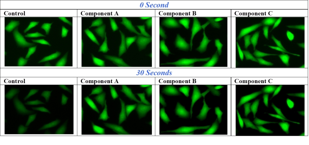
When exposed to excitation light, fluorescence intensity of dyes decreases due to their photooxidation or other photoreactions. There are very few fluorescent dyes that completely resist photobleaching. Frequently, when a section has been scanned repeatedly under strong excitation light, dyes could lose significant fluorescence signal before visual evaluation or photography can be accomplished. For examples, the photobleaching of fluoresceins (such as FITC-labeled antibodies) has become a major problem in fluorescence microscopy. In severe cases (such as phycoprotein-labeled bioconjugates), a fluorescence image of high resolution can not even be taken due to the extremely high photobleaching rate. Fluoroquest™ Anti-Fading Kit is to reduce the dye photobleaching rate, giving researchers longer observation time. The kit contains all the essential components that can be readily applied to imaging experiments. They are all premixed and ready-to-use solutions. This kit is designed for slide format while #20003 is designed for microplate format.
Platform
Fluorescence microscope
Excitation | Varies |
Emission | Varies |
Recommended plate | Black wall/clear bottom |
Components
Example protocol
AT A GLANCE
Protocol summary
- Prepare Samples (slides or microplate wells)
- Add a drop of a component and mount
- Examine the specimen under microscope
Important notes
These ready-to-use anti-fading reagents can be applied directly on the washed specimen. Although the reagents have been tested with lots of fixed samples, their optimal anti-fading efficiencies strongly depend on the properties of your samples. We suggest that you try more than one component for your imaging samples to get the ideal component. For example, one component may be more compatible with a fluorescent labeled antibody conjugate (or an enzyme substrate or a special mounting specimens that contain lipophilic plasma membrane stains like DiI) than another one.
SAMPLE EXPERIMENTAL PROTOCOL
Table 1. Recommendations for usage
Components | Recommendation |
Component A | Optimized for FITC and other fluorescein-based imaging experiments. |
Component B | Optimized for multiplexing imaging. In some cases, it enhances initial fluorescence intensity besides its anti-fading effect. |
Component C | Optimized for multiplexing imaging with minimal phototoxicity |
- Thaw all the kit components at room temperature, and keep from light.
- Remove any excess liquid from your specimen. Add a small drop of the selected component to the specimen. If the sample is on a slide or tissue culture dish, carefully place a coverslip on the drop, avoiding air bubbles. If the sample is on a coverslip, invert the coverslip on a clean glass slide. Remove any excess anti-fading component.
- The anti-fading reagents should be incubated for 2 hours to overnight. For long-term storage, seal the coverslip to the slide with nail polish or a plastic sealant. Mounted slides should be stored at 4ºC in the dark for optimum sample longevity. The fluorescence imaging would remain stable for many weeks. Samples can be imaged immediately after mounting. A typical image is shown in Figure 1.
Images

Figure 1. U2OS cells in a 96-well Costar black plate were loaded with 1 µM calcein, AM for 1 hour, fixing with 2% formaldehyde for 30 minutes. Anti-fading reagents were added to the samples after removing all the media. The FITC signals were compared at 0 and 30 seconds exposure time by using an Olympus fluorescence microscopy. The same exposure settings were used for all the images.
References
View all 18 references: Citation Explorer
On the mechanism of Trolox as antiblinking and antibleaching reagent
Authors: Cordes T, Vogelsang J, Tinnefeld P.
Journal: J Am Chem Soc (2009): 5018
Authors: Cordes T, Vogelsang J, Tinnefeld P.
Journal: J Am Chem Soc (2009): 5018
Relationship between DAPI-fluorescence fading and nuclear DNA content: An alternative method to DNA quantification
Authors: Gallardo-Escarate C, Alvarez-Borrego J, Von Br and E, Dupre E, Del Rio-Portilla MA.
Journal: Biol Res (2007): 29
Authors: Gallardo-Escarate C, Alvarez-Borrego J, Von Br and E, Dupre E, Del Rio-Portilla MA.
Journal: Biol Res (2007): 29
Strategies to improve photostabilities in ultrasensitive fluorescence spectroscopy
Authors: Widengren J, Chmyrov A, Eggeling C, Lofdahl PA, Seidel CA.
Journal: J Phys Chem A (2007): 429
Authors: Widengren J, Chmyrov A, Eggeling C, Lofdahl PA, Seidel CA.
Journal: J Phys Chem A (2007): 429
The reliability of long-term storage of direct immunofluorescent staining slides at room temperature
Authors: Dikicioglu E, Meteoglu I, Okyay P, Culhaci N, Kacar F.
Journal: J Cutan Pathol (2003): 430
Authors: Dikicioglu E, Meteoglu I, Okyay P, Culhaci N, Kacar F.
Journal: J Cutan Pathol (2003): 430
Comparative genomic hybridization technique
Authors: El-Rifai WE, Knuutila S.
Journal: Methods Mol Med (2001): 25
Authors: El-Rifai WE, Knuutila S.
Journal: Methods Mol Med (2001): 25
Fast-in situ hybridization and immunoenzymatic color pigment detection of mouse bone marrow micronucleus
Authors: Kolanko CJ, Pyle MD, Loats H, Parton J, Blakely WF, Nath J.
Journal: Biotech Histochem (1999): 111
Authors: Kolanko CJ, Pyle MD, Loats H, Parton J, Blakely WF, Nath J.
Journal: Biotech Histochem (1999): 111
Comparison of fixation protocols for adherent cultured cells applied to a GFP fusion protein of the epidermal growth factor receptor
Authors: Brock R, Hamelers IH, Jovin TM.
Journal: Cytometry (1999): 353
Authors: Brock R, Hamelers IH, Jovin TM.
Journal: Cytometry (1999): 353
Antifading agents for confocal fluorescence microscopy
Authors: Berrios M, Conlon KA, Colflesh DE.
Journal: Methods Enzymol (1999): 55
Authors: Berrios M, Conlon KA, Colflesh DE.
Journal: Methods Enzymol (1999): 55
Evaluation of five green fluorescence-emitting streptavidin-conjugated fluorochromes for use in immunofluorescence microscopy
Authors: Benchaib M, Delorme R, Pluvinage M, Bryon PA, Souchier C.
Journal: Histochem Cell Biol (1996): 253
Authors: Benchaib M, Delorme R, Pluvinage M, Bryon PA, Souchier C.
Journal: Histochem Cell Biol (1996): 253
Immunofluorescent artifacts due to the pH of antifading mounting media
Authors: Swartz DJ, Santi PA.
Journal: Biotechniques (1996): 398
Authors: Swartz DJ, Santi PA.
Journal: Biotechniques (1996): 398