logo
AAT Bioquest

Helixyte™ Green ssDNA reagent

The ssDNA dose response was measured with Helixyte™ Green ssDNA reagent in a 96-well solid black plate.
Ordering information
Price
Catalog Number
Unit Size
Quantity
Add to cart
Additional ordering information
Telephone1-800-990-8053
Fax1-800-609-2943
Emailsales@aatbio.com
InternationalSee distributors
Bulk requestInquire
Custom sizeInquire
ShippingStandard overnight for United States, inquire for international
Request quotation
Physical properties
SolventDMSO
Spectral properties
Excitation (nm)498
Emission (nm)519
Storage, safety and handling
H-phraseH303, H313, H333
Hazard symbolXN
Intended useResearch Use Only (RUO)
R-phraseR20, R21, R22
StorageFreeze (< -15 °C); Minimize light exposure
UNSPSC12171501

OverviewpdfSDSpdfProtocol


Excitation (nm)
498
Emission (nm)
519
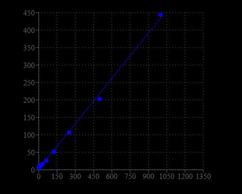
Synthetic oligonucleotides are used in several molecular biology techniques, such as DNA sequencing, site-directed mutagenesis, DNA amplification, and in situ hybridization. The most commonly used technique for measuring oligonucleotide and single-stranded DNA (ssDNA) concentration is the determination of absorbance at 260 nm (A260). However, absorbance methods suffer significant interferences resulting from various contaminants commonly found in nucleic acid preparations, including nucleotides, double-stranded nucleic acids, and proteins. Helixyte™ Green ssDNA Quantifying Reagent is an excellent alternative for quantifying ssDNA and oligonucleotides with significantly improved sensitivity and selectivity. It is a positively charged fluorescent probe that binds to the hydrophobic pockets of ssDNA and forms a highly luminescent complex through the synergistic actions of stacking, hydrophobic forces, hydrogen bonding, and electrostatic interactions. Helixyte™ Green ssDNA reagent has extremely low inherent fluorescence that is significantly enhanced upon binding to ssDNA. The increased sensitivity of Helixyte™ Green ssDNA over absorbance at 260 nm enables researchers to quantify as little as 100 pg/mL oligonucleotide or ssDNA (~500 pg/mL) with a standard spectrofluorometer and fluorescein excitation and emission wavelengths. As little as 1 ng/mL oligonucleotide or ssDNA can be detected with a fluorescence microplate reader. Helixyte™ Green ssDNA reagent has a wide linear range of detection ranging from 100 pg/mL to 1 μg/mL.

Platform


Fluorescence microplate reader

Excitation490 nm
Emission525 nm
Cutoff515 nm
Recommended plateSolid black

Example protocol


AT A GLANCE

Protocol summary
  1. Add 100 µL of ssDNA Standards or test samples
  2. Add 100 µL of Helixyte™ Green ssDNA working solution
  3. Incubate at RT for 5-10 minutes
  4. Monitor the fluorescence intensity at Ex/Em=490/525 nm 

Important
The following protocol is an example of quantifying the ssDNA using Helixyte™ Green ssDNA. Allow all the components to warm to room temperature before opening. No data are available on the mutagenicity or toxicity of Helixyte™ Green ssDNA stain. Because this reagent binds to nucleic acids, it should be treated as a potential mutagen and handled with appropriate care. The DMSO stock solution should be handled with particular caution as DMSO is known to facilitate the entry of organic molecules into tissues.

PREPARATION OF STANDARD SOLUTION

For convenience, use the Serial Dilution Planner:
https://www.aatbio.com/tools/serial-dilution/17620


ssDNA Standard solution
Add 10 uL of 100 ug/mL ssDNA Standard solution (Not provided) to 190 uL of buffer of your choice to get a 5 ug/mL standards solution, then perform 1:3 dilutions to obtain serially diluted ssDNA standards (SS2-SS7).

PREPARATION OF WORKING SOLUTION

Helixyte™ Green ssDNA working solution
Prepare the Helixyte™ Green ssDNA working solution by adding 100 μL of Helixyte™ Green ssDNA reagent into 10 mL of buffer of your choice. Protect the working solution from light by covering it with foil or placing it in the dark.
Note     It’s recommended to prepare the working solution in a plastic container rather than a glass container, as the dye may adsorb to the glass surface. For best results, this solution should be used within a few hours after the dilution.
Note     10 mL of working solution is enough for one 96-well plate.
Note     10 mM Tris-HCl (pH 8.0) with 0.1 mM EDTA can be used to make working solution and standards. 

SAMPLE EXPERIMENTAL PROTOCOL

Table 1.The layout of ssDNA Standards and test samples in a solid black 96-well microplate. SS= ssDNA Standards (SS1 - SS7, 1667 to 2.3 ng/mL); BL=Blank Control; TS=Test Samples
BL BL TS TS
SS1 SS1
SS2 SS2
SS3 SS3    
SS4 SS4    
SS5 SS5    
SS6 SS6    
SS7 SS7    
Table 2.The reagent composition for each well.
WellVolumeReagent
SS1-SS7100 µLSerial dilutions (1667 to 2.3 ng/mL)
BL100 µLBuffer of your choice
TS100 µLSample
  1. Prepare ssDNA Standards (NS), blank controls (BL), and test samples (TS) according to the layout provided in Tables 1 and 2. For a 384-well plate, use 25 µL of reagent per well instead of 100 µL.
  2. Add 100 µL of the Helixyte™ Green ssDNA working solution to each well of ssDNA Standards, blank control, and test samples to make the assay volume of 200 µL/well. For a 384-well plate, add 25 µL of the Helixyte™ Green ssDNA working solution into each well instead, to get a total volume of 50 µL/well.
  3. Incubate the reaction at room temperature for 5 to 10 minutes, protected from light.
  4. Monitor the fluorescence increase with a fluorescence microplate reader at Ex/Em = 490/525 nm (cut off at 515 nm). 

Spectrum


Open in Advanced Spectrum Viewer
spectrum

Spectral properties

Excitation (nm)498
Emission (nm)519

Images


References


View all 50 references: Citation Explorer
Dual-Channel Logic Gates Operating on the Chemopalette ssDNA-Ag NCs/GO Nanocomposites.
Authors: Feng, Da-Qian and Liu, Guoliang
Journal: Analytical chemistry (2021)
Biotechnological production of ssDNA with DNA-hydrolyzing deoxyribozymes.
Authors: Liu, Jin and Gu, Hongzhou
Journal: STAR protocols (2021): 100531
Molecular underpinnings of ssDNA specificity by Rep HUH-endonucleases and implications for HUH-tag multiplexing and engineering.
Authors: Tompkins, Kassidy J and Houtti, Mo and Litzau, Lauren A and Aird, Eric J and Everett, Blake A and Nelson, Andrew T and Pornschloegl, Leland and Limón-Swanson, Lidia K and Evans, Robert L and Evans, Karen and Shi, Ke and Aihara, Hideki and Gordon, Wendy R
Journal: Nucleic acids research (2021): 1046-1064
Efficient influence of ssDNA virus PCV2 replication by CRISPR/Cas9 targeting of the viral genome.
Authors: Shi, Jianli and Zheng, Shuxuan and Wu, Xiaoyan and Peng, Zhe and Li, Chen and Wang, Shuo and Xin, Changxun and Xu, Shaojian and Li, Jun
Journal: Molecular immunology (2021): 63-66
Translocation of flexible and tensioned ssDNA through in silico designed hydrophobic nanopores with two constrictions.
Authors: Rattu, Punam and Belzunces, Bastien and Haynes, Taylor and Skylaris, Chris-Kriton and Khalid, Syma
Journal: Nanoscale (2021): 1673-1679
Intra-host evolution of the ssDNA virus tomato severe rugose virus (ToSRV).
Authors: Pinto, Vitor Batista and Quadros, Ayane Fernanda Ferreira and Godinho, Márcio Tadeu and Silva, José Cleydson and Alfenas-Zerbini, Poliane and Zerbini, F Murilo
Journal: Virus research (2021): 198234
A Synergistic Coreactant for Single-Cell Electrochemiluminescence Imaging: Guanine-Rich ssDNA-Loaded High-Index Faceted Gold Nanoflowers.
Authors: Chen, Ying and Gou, Xiaodan and Ma, Cheng and Jiang, Dechen and Zhu, Jun-Jie
Journal: Analytical chemistry (2021): 7682-7689
Single-Stranded DNA Curtains for Single-Molecule Visualization of Rad51-ssDNA Filament Dynamics.
Authors: Roy, Upasana and Greene, Eric C
Journal: Methods in molecular biology (Clifton, N.J.) (2021): 193-207
Nitrogen-doped carbon dots aid in the separation of ssDNA molecules of different length by capillary transient isotachophoresis (ctITP) with laser-induced fluorescence (LIF) detection.
Authors: Roy, Debashish and Colyer, Christa L
Journal: Journal of chromatography. A (2021): 461990
Selection and identification of an ssDNA aptamer to NB4 cell.
Authors: Zhang, Xian-Hui and Wang, Wei and Chen, Xin
Journal: Journal of clinical laboratory analysis (2021): e23718