iFluor® 568 goat anti-rabbit IgG (H+L) *Cross Adsorbed*
iFluor® 568 is a bright red fluorescent dye. iFluor® 568-labeled anti-IgG conjugates exhibit bright fluorescence signal and good photostability. Used for stable signal generation in imaging and flow cytometry, the fluorescence intensity of iFluor® 568 conjugates is pH-insensitive from pH 4 to pH 11. The iFluor® 568-labeled antibody conjugates can be well excited with Krypton ion laser (~568 nm). iFluor® 568 family has the spectral properties essentially identical to those of Alexa Fluor® 568. Under the same conditions we tested, iFluor® 568 antibody conjugates are brighter and more photostable than the corresponding Alexa Fluo® 568. These spectral and labeling characteristics make the iFluor® 568 dye family a superior alternative to Alexa Fluor® 568. In addition, iFluor® 568 secondary antibody conjugates give higher signal/background ratios than the corresponding Alexa Fluor® 568-labeled conjugates.
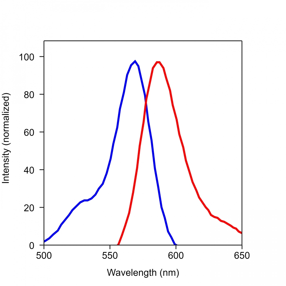
Spectrum
Open in Advanced Spectrum Viewer
Product family
Name | Excitation (nm) | Emission (nm) | Extinction coefficient (cm -1 M -1) | Quantum yield | Correction Factor (260 nm) | Correction Factor (280 nm) |
iFluor® 350 goat anti-rabbit IgG (H+L) | 345 | 450 | 200001 | 0.951 | 0.83 | 0.23 |
iFluor® 405 goat anti-rabbit IgG (H+L) | 403 | 427 | 370001 | 0.911 | 0.48 | 0.77 |
iFluor® 488 goat anti-rabbit IgG (H+L) | 491 | 516 | 750001 | 0.91 | 0.21 | 0.11 |
iFluor® 514 goat anti-rabbit IgG (H+L) | 511 | 527 | 750001 | 0.831 | 0.265 | 0.116 |
iFluor® 532 goat anti-rabbit IgG (H+L) | 537 | 560 | 900001 | 0.681 | 0.26 | 0.16 |
iFluor® 555 goat anti-rabbit IgG (H+L) | 557 | 570 | 1000001 | 0.641 | 0.23 | 0.14 |
iFluor® 594 goat anti-rabbit IgG (H+L) | 587 | 603 | 2000001 | 0.531 | 0.05 | 0.04 |
iFluor® 633 goat anti-rabbit IgG (H+L) | 640 | 654 | 2500001 | 0.291 | 0.062 | 0.044 |
iFluor® 647 goat anti-rabbit IgG (H+L) | 656 | 670 | 2500001 | 0.251 | 0.03 | 0.03 |
Show More (31) |
Citations
View all 4 citations: Citation Explorer
Overexpression of CXCR2 predicts poor prognosis in patients with colorectal cancer.
Authors: Zhao, Jingkun and Ou, Baochi and Feng, Hao and Wang, Puxiongzhi and Yin, Shuai and Zhu, Congcong and Wang, Shenjie and Chen, Chun and Zheng, Minhua and Zong, Yaping and others, undefined
Journal: Oncotarget (2017)
Authors: Zhao, Jingkun and Ou, Baochi and Feng, Hao and Wang, Puxiongzhi and Yin, Shuai and Zhu, Congcong and Wang, Shenjie and Chen, Chun and Zheng, Minhua and Zong, Yaping and others, undefined
Journal: Oncotarget (2017)
Cadherin-12 enhances proliferation in colorectal cancer cells and increases progression by promoting EMT
Authors: Ma, Junjun and Zhao, Jingkun and Lu, Jun and Wang, Puxiongzhi and Feng, Hao and Zong, Yaping and Ou, Baochi and Zheng, Minhua and Lu, Aiguo
Journal: Tumor Biology (2016): 1--12
Authors: Ma, Junjun and Zhao, Jingkun and Lu, Jun and Wang, Puxiongzhi and Feng, Hao and Zong, Yaping and Ou, Baochi and Zheng, Minhua and Lu, Aiguo
Journal: Tumor Biology (2016): 1--12
Transplantation of RADA16-BDNF peptide scaffold with human umbilical cord mesenchymal stem cells forced with CXCR4 and activated astrocytes for repair of traumatic brain injury
Authors: Shi, W and Huang, CJ and Xu, XD and Jin, GH and Huang, RQ and Huang, JF and Chen, YN and Ju, SQ and Wang, Y and Shi, YW and others, undefined
Journal: Acta Biomaterialia (2016): 247--261
Authors: Shi, W and Huang, CJ and Xu, XD and Jin, GH and Huang, RQ and Huang, JF and Chen, YN and Ju, SQ and Wang, Y and Shi, YW and others, undefined
Journal: Acta Biomaterialia (2016): 247--261
The migration and differentiation of hUC-MSCsCXCR4/GFP encapsulated in BDNF/chitosan scaffolds for brain tissue engineering
Authors: Huang, Chuanjun and Zhao, Longxiang and Gu, Jun and Nie, Dekang and Chen, Yinan and Zuo, Hao and Huan, Wei and Shi, Jinlong and Chen, Jian and Shi, Wei
Journal: Biomedical Materials (2016): 035004
Authors: Huang, Chuanjun and Zhao, Longxiang and Gu, Jun and Nie, Dekang and Chen, Yinan and Zuo, Hao and Huan, Wei and Shi, Jinlong and Chen, Jian and Shi, Wei
Journal: Biomedical Materials (2016): 035004
References
View all 191 references: Citation Explorer
Antiprothrombin antibodies in a patient with secondary antiphospholipid syndrome and bleeding
Authors: Gonzalez Leon R, Garcia Hern and ez FJ, Castillo Palma MJ, Sanchez Roman J.
Journal: Med Clin (Barc) (2011): 668
Authors: Gonzalez Leon R, Garcia Hern and ez FJ, Castillo Palma MJ, Sanchez Roman J.
Journal: Med Clin (Barc) (2011): 668
Assessment of EGFR/HER2 dimerization by FRET-FLIM utilizing Alexa-conjugated secondary antibodies in relation to targeted therapies in cancers
Authors: Waterhouse BR, Gijsen M, Barber PR, Tullis ID, Vojnovic B, Kong A.
Journal: Oncotarget (2011): 728
Authors: Waterhouse BR, Gijsen M, Barber PR, Tullis ID, Vojnovic B, Kong A.
Journal: Oncotarget (2011): 728
Falsely elevated tacrolimus levels caused by immunoassay interference secondary to beta-galactosidase antibodies in an infected liver transplant recipient
Authors: Knorr JP, Grewal KS, Balasubramanian M, Young N, Zaki R, Khanmoradi K, Araya V, Ortiz J.
Journal: Pharmacotherapy (2010): 954
Authors: Knorr JP, Grewal KS, Balasubramanian M, Young N, Zaki R, Khanmoradi K, Araya V, Ortiz J.
Journal: Pharmacotherapy (2010): 954
Prevalence of anti-Epstein-Barr virus antibodies in children and adolescents with secondary immunodeficiency
Authors: Buckova A., undefined
Journal: Epidemiol Mikrobiol Imunol (2010): 133
Authors: Buckova A., undefined
Journal: Epidemiol Mikrobiol Imunol (2010): 133
Analysis of the effectiveness of reused primary and secondary antibodies in Western blotting analysis
Authors: Boonrod K, Roth B, Leong Ngar S, Krczal G.
Journal: Anal Biochem (2010): 124
Authors: Boonrod K, Roth B, Leong Ngar S, Krczal G.
Journal: Anal Biochem (2010): 124
Page updated on December 11, 2024