logo
AAT Bioquest

iFluor® 790 goat anti-mouse IgG (H+L) *Cross Adsorbed*

AAT Bioquest's iFluor® dyes are optimized for labeling proteins, in particular, antibodies. These dyes are bright, photostable and have minimal quenching on proteins. They can be well excited by the major laser lines of fluorescence instruments (e.g., 350, 405, 488, 555 and 633 nm). iFluor® 790 goat anti-mouse IgG (H+L) conjugate has IR fluorescence excitation and emission maxima of ~780 nm and ~810 nm respectively. These spectral characteristics make them an excellent alternative to IRDye® 800 goat anti-mouse IgG (H+L) conjugate (IRDye® is the trademark of Li-COR).
Spectrum

Product family

NameExcitation (nm)Emission (nm)Extinction coefficient (cm -1 M -1)Quantum yieldCorrection Factor (260 nm)Correction Factor (280 nm)
iFluor® 350 goat anti-mouse IgG (H+L)3454502000010.9510.830.23
iFluor® 405 goat anti-mouse IgG (H+L)4034273700010.9110.480.77
iFluor® 488 goat anti-mouse IgG (H+L)4915167500010.910.210.11
iFluor® 514 goat anti-mouse IgG (H+L)5115277500010.8310.2650.116
iFluor® 532 goat anti-mouse IgG (H+L)5375609000010.6810.260.16
iFluor® 555 goat anti-mouse IgG (H+L)55757010000010.6410.230.14
iFluor® 594 goat anti-mouse IgG (H+L)58760320000010.5310.050.04
iFluor® 633 goat anti-mouse IgG (H+L)64065425000010.2910.0620.044
iFluor® 647 goat anti-mouse IgG (H+L)65667025000010.2510.030.03
iFluor® 680 goat anti-mouse IgG (H+L)68470122000010.2310.0970.094
iFluor® 700 goat anti-mouse IgG (H+L)69071322000010.2310.090.04
iFluor® 750 goat anti-mouse IgG (H+L)75777927500010.1210.0440.039
iFluor® 350 goat anti-mouse IgG (H+L) *Cross Adsorbed*3454502000010.9510.830.23
iFluor® 405 goat anti-mouse IgG (H+L) *Cross Adsorbed*4034273700010.9110.480.77
iFluor® 488 goat anti-mouse IgG (H+L) *Cross Adsorbed*4915167500010.910.210.11
iFluor® 514 goat anti-mouse IgG (H+L) *Cross Adsorbed*5115277500010.8310.2650.116
iFluor® 532 goat anti-mouse IgG (H+L) *Cross Adsorbed*5375609000010.6810.260.16
iFluor® 555 goat anti-mouse IgG (H+L) *Cross Adsorbed*55757010000010.6410.230.14
iFluor® 594 goat anti-mouse IgG (H+L) *Cross Adsorbed*58760320000010.5310.050.04
iFluor® 633 goat anti-mouse IgG (H+L) *Cross Adsorbed*64065425000010.2910.0620.044
iFluor® 647 goat anti-mouse IgG (H+L) *Cross Adsorbed*65667025000010.2510.030.03
iFluor® 680 goat anti-mouse IgG (H+L) *Cross Adsorbed*68470122000010.2310.0970.094
iFluor® 700 goat anti-mouse IgG (H+L) *Cross Adsorbed*69071322000010.2310.090.04
iFluor® 750 goat anti-mouse IgG (H+L) *Cross Adsorbed*75777927500010.1210.0440.039
iFluor® 790 goat anti-rabbit IgG (H+L)78781225000010.1310.10.09
iFluor® 790 goat anti-rabbit IgG (H+L) *Cross Adsorbed*78781225000010.1310.10.09
iFluor® 546 goat anti-mouse IgG (H+L)54155710000010.6710.250.15
iFluor® 546 goat anti-mouse IgG (H+L) *Cross Adsorbed*54155710000010.6710.250.15
iFluor® 568 goat anti-mouse IgG (H+L)56858710000010.5710.340.15
iFluor® 568 goat anti-mouse IgG (H+L) *Cross Adsorbed*56858710000010.5710.340.15
iFluor® 800 goat anti-mouse IgG (H+L) 80182025000010.1110.030.08
iFluor® 800 goat anti-mouse IgG (H+L) *Cross Adsorbed*80182025000010.1110.030.08
iFluor® 810 goat anti-mouse IgG (H+L) 81182225000010.0510.090.15
iFluor® 810 goat anti-mouse IgG (H+L) *Cross Adsorbed*81182225000010.0510.090.15
iFluor® 820 goat anti-mouse IgG (H+L)82285025000010.110.16
iFluor® 820 goat anti-mouse IgG (H+L) *Cross Adsorbed*82285025000010.110.16
iFluor® 840 goat anti-mouse IgG (H+L)8368792000001-0.20.09
iFluor® 840 goat anti-mouse IgG (H+L) *Cross Adsorbed*8368792000001-0.20.09
iFluor® 860 goat anti-mouse IgG (H+L)85387825000010.10.14
iFluor® 860 goat anti-mouse IgG (H+L) *Cross Adsorbed*85387825000010.10.14
Show More (31)

Citations

View all 8 citations: Citation Explorer
Electronic-Resonance Coherent Anti-Stokes Raman Scattering Spectroscopy and Microscopy
Authors: Tang, Qi and Li, Baoguo and Wang, Jianjun and Liu, Yufeng and Pinkas, Iddo and Rigneault, Herv{\'e} and Oron, Dan and Ren, Liqing
Journal: ACS Photonics (2024)
Concentrated growth factor matrices prepared using silica-coated plastic tubes are distinguishable from those prepared using glass tubes in platelet distribution: application of a novel near-infrared imaging-based, quantitative technique
Authors: Yamaguchi, Sadahiro and Aizawa, Hachidai and Sato, Atsushi and Tsujino, Tetsuhiro and Isobe, Kazushige and Kitamura, Yutaka and Watanabe, Taisuke and Okudera, Hajime and Mour{\~a}o, Carlos Fernando and Kawase, Tomoyuki
Journal: Frontiers in Bioengineering and Biotechnology (2020): 600
Quantitative near-infrared imaging of platelets in platelet-rich fibrin (PRF) matrices: comparative analysis of bio-PRF, leukocyte-rich PRF, advanced-PRF and concentrated growth factors
Authors: Aizawa, Hachidai and Tsujino, Tetsuhiro and Watanabe, Taisuke and Isobe, Kazushige and Kitamura, Yutaka and Sato, Atsushi and Yamaguchi, Sadahiro and Okudera, Hajime and Okuda, Kazuhiro and Kawase, Tomoyuki
Journal: International Journal of Molecular Sciences (2020): 4426
Distribution and quantification of activated platelets in platelet-rich fibrin matrices
Authors: Sato, Atsushi and Kawabata, Hideo and Aizawa, Hachidai and Tsujino, Tetsuhiro and Isobe, Kazushige and Watanabe, Taisuke and Kitamura, Yutaka and Miron, Richard J and Kawase, Tomoyuki
Journal: Platelets (2020): 1--6
Overexpression of CXCR2 predicts poor prognosis in patients with colorectal cancer.
Authors: Zhao, Jingkun and Ou, Baochi and Feng, Hao and Wang, Puxiongzhi and Yin, Shuai and Zhu, Congcong and Wang, Shenjie and Chen, Chun and Zheng, Minhua and Zong, Yaping and others, undefined
Journal: Oncotarget (2017)

References

View all 191 references: Citation Explorer
Antiprothrombin antibodies in a patient with secondary antiphospholipid syndrome and bleeding
Authors: Gonzalez Leon R, Garcia Hern and ez FJ, Castillo Palma MJ, Sanchez Roman J.
Journal: Med Clin (Barc) (2011): 668
Assessment of EGFR/HER2 dimerization by FRET-FLIM utilizing Alexa-conjugated secondary antibodies in relation to targeted therapies in cancers
Authors: Waterhouse BR, Gijsen M, Barber PR, Tullis ID, Vojnovic B, Kong A.
Journal: Oncotarget (2011): 728
Falsely elevated tacrolimus levels caused by immunoassay interference secondary to beta-galactosidase antibodies in an infected liver transplant recipient
Authors: Knorr JP, Grewal KS, Balasubramanian M, Young N, Zaki R, Khanmoradi K, Araya V, Ortiz J.
Journal: Pharmacotherapy (2010): 954
Three siblings with self-resolving congenital hyperthyrotropinaemia secondary to thyrotropin receptor blocking antibodies
Authors: Azzopardi P, Forrester M, Ehtisham S.
Journal: J Paediatr Child Health (2010): 439
Nanosilver-doped DNA polyion complex membrane for electrochemical immunoassay of carcinoembryonic antigen using nanogold-labeled secondary antibodies
Authors: Wu W, Yi P, He P, Jing T, Liao K, Yang K, Wang H.
Journal: Anal Chim Acta (2010): 126
Page updated on September 19, 2024

Ordering information

Price
Unit size
200 ug
1 mg
Catalog Number
Quantity
Add to cart

Additional ordering information

Telephone1-800-990-8053
Fax1-800-609-2943
Emailsales@aatbio.com
InternationalSee distributors
Bulk requestInquire
Custom sizeInquire
Technical SupportContact us
Purchase orderSend to sales@aatbio.com
ShippingStandard overnight for United States, inquire for international
Request quotation

Physical properties

Molecular weight

~150000

Solvent

Water

Spectral properties

Correction Factor (260 nm)

0.1

Correction Factor (280 nm)

0.09

Extinction coefficient (cm -1 M -1)

2500001

Excitation (nm)

787

Emission (nm)

812

Quantum yield

0.131

Storage, safety and handling

H-phraseH303, H313, H333
Hazard symbolXN
Intended useResearch Use Only (RUO)
R-phraseR20, R21, R22

Storage

Freeze (< -15 °C); Minimize light exposure
UNSPSC12171501
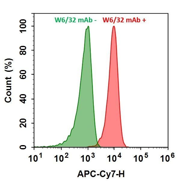
HL-60 cells were incubated with (Red, +) or without (Green, -) Anti-human HLA-ABC (W6/32 mAb), followed by iFluor® 790 goat anti-mouse IgG conjugate. The fluorescence signal was monitored using ACEA NovoCyte flow cytometer in APC-Cy7 channel.