logo
AAT Bioquest

MMP Green™ substrate

The internally quenched FRET peptide substrate is digested by a protease to generate the highly fluorescent peptide fragment. The fluorescence increase is proportional to the protease activity.
Ordering information
Price
Catalog Number
Unit Size
Quantity
Add to cart
Additional ordering information
Telephone1-800-990-8053
Fax1-800-609-2943
Emailsales@aatbio.com
InternationalSee distributors
Bulk requestInquire
Custom sizeInquire
ShippingStandard overnight for United States, inquire for international
Request quotation
Physical properties
Molecular weight~2000
SolventDMSO
Spectral properties
Excitation (nm)494
Emission (nm)515
Storage, safety and handling
H-phraseH303, H313, H333
Hazard symbolXN
Intended useResearch Use Only (RUO)
R-phraseR20, R21, R22
StorageFreeze (< -15 °C); Minimize light exposure
UNSPSC12352200
Alternative formats
MMP Red™ substrate

OverviewpdfSDSpdfProtocol


See also: Proteases
Molecular weight
~2000
Excitation (nm)
494
Emission (nm)
515
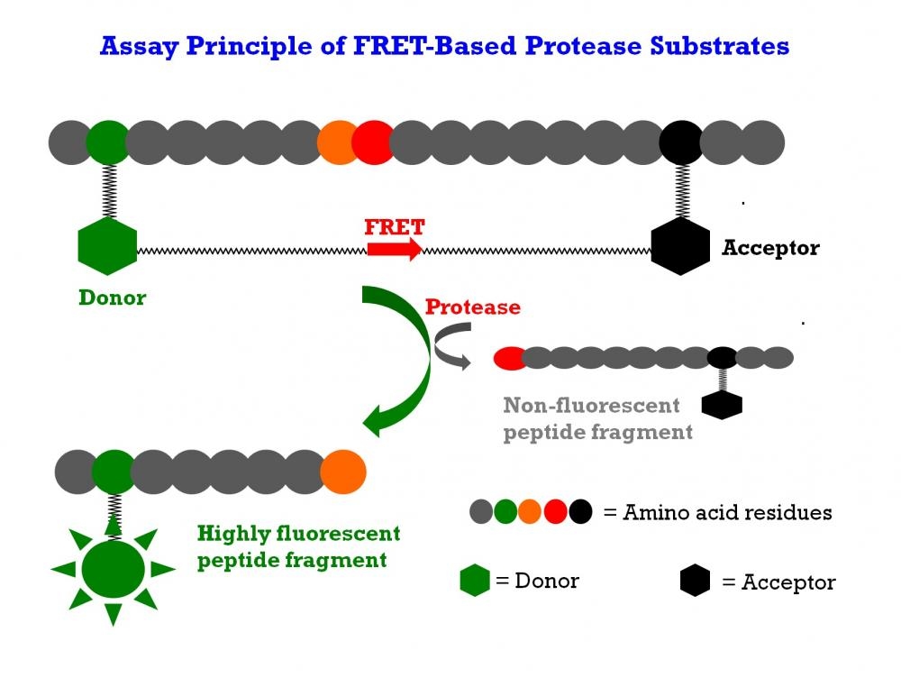
The matrix metalloproteinases (MMPs) constitute a family of zinc-dependent endopeptidases that function within the extracellular matrix. These enzymes are responsible for the breakdown of connective tissues and are important in bone remodeling, the menstrual cycle and repair of tissue damage. While the exact contribution of MMPs to certain pathological processes is difficult to assess, MMPs appear to play a key role in the development of arthritis as well as in the invasion and metastasis of cancer. MMPs tend to have multiple substrates, with most family members having the ability to degrade different types of collagen along with elastin, gelatin and fibronectin. It is quite difficult to find a substrate that is selective to a single MMP enzyme. This FRET substrate is designed to monitor the general activity of a MMP enzyme. It can also be used to screening MMP inhibitors when a purified MMP enzyme is used. This FRET substrate is based on our TF2/TQ2 FRET pair. Upon MMP hydrolysis the fluorescence of MMP Green™ FRET peptide substrate is increased since the TF2/TQ2 FRET pair is separated. The fluorescence increase is proportional to the MMP enzyme activities.

Platform


Fluorescence microplate reader

Excitation490 nm
Emission525 nm
Cutoff515 nm
Recommended plateSolid black

Example protocol


AT A GLANCE

Protocol summary
  1. Add appropriate controls or test samples with or without APMA (50 µL)
  2. Pre-incubate for 10 - 15 minutes
  3. Add MMP Green™ substrate working solution (50 µL)
  4. Skip incubation step for kinetic reading or incubate for 30 to 60 minutes for end point reading
  5. Monitor fluorescence intensity at Ex/Em = 490/525 nm 
Important      Thaw the reagents includig the substrate at room temperature before starting the experiment. Prepare MMPs containing biological samples as desired.

PREPARATION OF STOCK SOLUTIONS

Unless otherwise noted, all unused stock solutions should be divided into single-use aliquots and stored at -20 °C after preparation. Avoid repeated freeze-thaw cycles.

MMP Green™ Substrate stock solution
For Cat# 13520, prepare 1- 10 mg/mL stock solution by adding appropriate amount of DMSO to MMP Green™ substrate vial.
Note     Store stock solution in single used aliquots and protect from light.

PREPARATION OF WORKING SOLUTION

MMP Green™ Substrate working solution
For Cat# 13520, prepare 20-100 µg/mL MMP Green™ Substrate working solution in buffer of your choice. For Cat# 13519, Add 50 µL of MMP Green™ solution (vial provided) into 5 mL of buffer of your choice.

SAMPLE EXPERIMENTAL PROTOCOL

Table 1.Protocols for MMP activation.
MMPsActivated by treating with
MMP-1 (collagenase)1 mM APMA (diluted component C) at 37 °C for 3 hr
MMP-2 (gelatinase)1 mM APMA (diluted component C) at 37 °C for 1 hr
MMP-3 (stromelysin)1 mM APMA (diluted component C) at 37 °C for 24 hr
MMP-7 (matrilysin, PUMP-1)1 mM APMA (diluted component C) at 37 °C for 20 min - 1 hr
MMP-8 (neutrophil collagenase)1 mM APMA (diluted component C) at 37 °C for 1 hr
MMP-9 (92 kDa gelatinase)1 mM APMA (diluted component C) at 37 °C for 2 h
MMP-10 (stromelysin-2)1 mM APMA (diluted component C) at 37 °C for 24 hr
MMP-11 (stromelysin-3)Already in active form. No APMA treatment is necessary
MMP-12 (macrophage elastase)1 mM APMA (diluted component C) at 37 °C for 2 hr
MMP-13 (collagenase-3)1 mM APMA (diluted component C) at 37°C for 40 min
MMP-141 mM APMA (diluted component C) at 37°C for 2 - 3 hr
  1. Prepare MMPs containing biological samples as desired.
  2. To activate pro-MMPs, add 2 mM APMA working solution (2X) to the plate.
    Note     APMA belongs to organic mercury. Handle with care! Dispose it according to local regulations. Next, incubate the MMP containing-samples or purified MMPs with equal volume of 2 mM APMA working solution (2X). Refer to Table 1 for incubation time. Activate MMPs immediately before the experiment.
    Note     Keep enzyme-containing samples on ice. Avoid vigorously vortexing the enzyme. Prolonged storage of the activated enzyme will deactivate the enzyme. For enzyme activation, it is preferably activated at higher protein concentration. After activation, you may further dilute the enzyme.
  3. Prepare controls and test samples (TS) with 50 µL per well. For a 384-well plate, use 20 µL of reagent per well instead of 50 µL.
  4. Pre-incubate the plate at a desired temperature for the enzyme reaction (e.g. 25 °C or 37 °C) for 10 - 15 minutes if you are screening MMPs inhibitors.
  5. Add 50 µL (96-well) or 20 µL (384-well) of MMP Green™ substrate working solution to the sample and control wells of the assay plate. Mix the reagents well.
  6. Monitor the fluorescence intensity with a fluorescence plate reader at Ex/Em = 490/525 nm. For kinetic reading: Immediately start measuring fluorescence intensity and continuously record data every 5 minutes for 30 to 60 minutes. For end-point reading: Incubate the reaction at room temperature for 30 to 60 minutes, kept from light if possible. Mix the reagents well, and then measure the fluorescence intensity. 

Spectrum


Open in Advanced Spectrum Viewer
spectrum

Spectral properties

Excitation (nm)494
Emission (nm)515

Product Family


NameExcitation (nm)Emission (nm)
MMP Red™ substrate545572

Images


Citations


View all 11 citations: Citation Explorer
Octenidine-based hydrogel shows anti-inflammatory and protease-inhibitory capacities in wounded human skin
Authors: Seiser, Saskia and Janker, Lukas and Zila, Nina and Mildner, Michael and Rakita, Ana and Matiasek, Johannes and Bileck, Andrea and Gerner, Christopher and Paulitschke, Verena and Elbe-B{\"u}rger, Adelheid
Journal: Scientific Reports (2021): 1--13
Zinc ions regulate opening of tight junction favouring efflux of macromolecules via the GSK3&beta;/snail-mediated pathway
Authors: Xiao, Ruyue and Yuan, Lan and He, Weijiang and Yang, Xiaoda
Journal: Metallomics (2018)
Connexin 43 Upregulation by Dioscin Inhibits Melanoma Progression via Suppressing Malignancy and Inducing M1 Polarization
Authors: Kou, Yu and Ji, Liyan and Wang, Haojia and Wang, Wensheng and Zheng, Hongming and Zou, Juan and Liu, Linxin and Qi, Xiaoxiao and Liu, Zhongqiu and Du, Biaoyan and others, undefined
Journal: International Journal of Cancer (2017)
DACT2, an epigenetic stimulator, exerts dual efficacy for colorectal cancer prevention and treatment
Authors: Lu, Linlin and Wang, Ying and Ou, Rilan and Feng, Qian and Ji, Liyan and Zheng, Hongming and Guo, Yue and Qi, Xiaoxiao and Kong, Ah-Ng Tony and Liu, Zhongqiu
Journal: Pharmacological research (2017)
Probing Cell Adhesion Profiles with a Microscale Adhesive Choice Assay
Authors: Kittur, Harsha and Tay, Andy and Hua, Avery and Yu, Min and Di Carlo, Dino
Journal: Biophysical Journal (2017): 1858--1867
Inhibition of BET bromodomain attenuates angiotensin II induced abdominal aortic aneurysm in ApoE-/- mice
Authors: Duan, Qiong and Mao, Xiaoxiao and Liao, Chaonan and Zhou, Haoyang and Sun, Zelin and Deng, Xu and Hu, Qiuning and Qi, Jun and Zhang, Guogang and Huang, He and others, undefined
Journal: International Journal of Cardiology (2016): 428--432
Tissue inhibitor of metalloproteinase 1 influences vascular adaptations to chronic alterations in blood flow
Authors: M, undefined and el, Erin R and Uchida, Cass and ra , undefined and Nwadozi, Emmanuel and Makki, Armin and Haas, Tara L
Journal: Journal of Cellular Physiology (2016)
Matrix Remodeling Maintains Embryonic Stem Cell Self-Renewal by Activating Stat3
Authors: Przybyla, Laralynne M and Theunissen, Thorold W and Jaenisch, Rudolf and Voldman, Joel
Journal: Stem Cells (2013): 1097--1106
&beta;-adrenoceptors are upregulated in human melanoma and their activation releases pro-tumorigenic cytokines and metalloproteases in melanoma cell lines
Authors: Moretti, Silvia and Massi, Daniela and Farini, Valentina and Baroni, Gianna and Parri, Matteo and Innocenti, Stefania and Cecchi, Roberto and Chiarugi, Paola
Journal: Laboratory Investigation (2013): 279--290
Identification and functional validation of CDH11, PCSK6 and SH3GL3 as novel glioma invasion-associated candidate genes
Authors: Delic, S and Lottmann, N and Jetschke, K and Reifenberger, G and Riemenschneider, Markus J
Journal: Neuropathology and applied neurobiology (2012): 201--212