logo
AAT Bioquest

Protonex™ Green 500-PEG12, SE

The pH dependent Emission spectra of Protonex™ Green 500-PEG12.
Ordering information
Price
Catalog Number
Unit Size
Quantity
Add to cart
Additional ordering information
Telephone1-800-990-8053
Fax1-800-609-2943
Emailsales@aatbio.com
InternationalSee distributors
Bulk requestInquire
Custom sizeInquire
ShippingStandard overnight for United States, inquire for international
Request quotation
Physical properties
Molecular weight1139.26
SolventDMSO
Spectral properties
Excitation (nm)453
Emission (nm)522
Storage, safety and handling
H-phraseH303, H313, H333
Hazard symbolXN
Intended useResearch Use Only (RUO)
R-phraseR20, R21, R22
StorageFreeze (< -15 °C); Minimize light exposure

OverviewpdfSDSpdfProtocol


Molecular weight
1139.26
Excitation (nm)
453
Emission (nm)
522
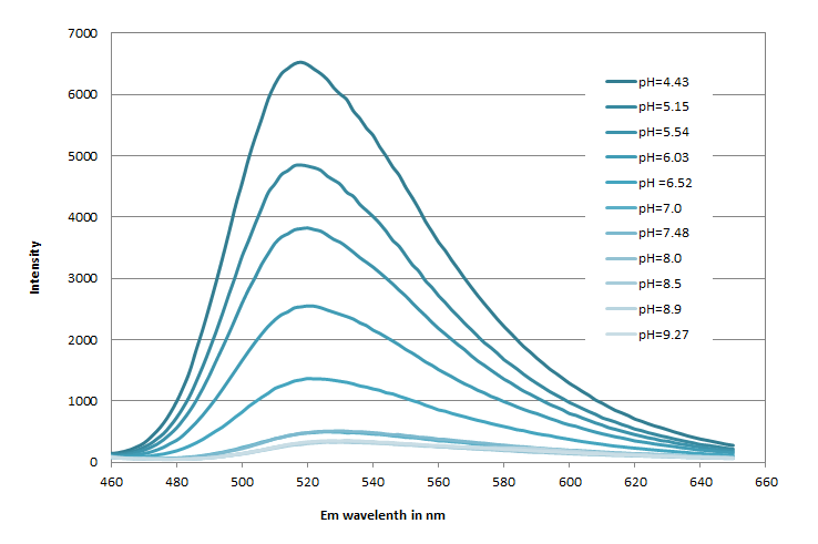
The amino-reactive Protonex™ Green 500 SE (#21216) was developed for preparing Protonex™ Green 500 conjugates as fluorescent pH probes. However, the utility of Protonex™ Green 500 SE was limited by its poor solubility. Protonex™ Green 500-PEG12 SE (#21219) has been developed to significantly improve the poor solubility of Protonex™ Green 500 SE (#21216) while maintaining the excellent spectral and pH properties of Protonex™ Green 500. They have identical spectra and pH profile while Green 500-PEG12 has significantly improved solubility. Protonex™ Green 500 dye demonstrated pH-dependent fluorescence. Unlike most of the existing fluorescent dyes that are more fluorescent at higher pH, acidic conditions enhance the fluorescence of Protonex™ Green 500 dye, making it an excellent acidotropic fluorescent probe. The fluorescence of Protonex™ Green 500 dye increases as pH decreases from neutral to the acidic. The lack of fluorescence outside the cell eliminates the wash steps. Protonex™ Green dye provides a powerful tool to monitor acidic cell compartments such as endosomes and lysosomes. Protonex™ Green dye is non-fluorescent outside the cells but fluoresces brightly green in acidic compartments (such as phagosomes, lysosomes and endosomes). This Protonex™ Green enables the specific detection of cellular acidic compartments with reduced signal variability and improved accuracy for imaging or flow applications. Protonex™ Green has the spectral properties similar to those of FITC, making the common filter set of FITC readily available to the assays of Protonex™ Green.

Example protocol


PREPARATION OF STOCK SOLUTIONS

Unless otherwise noted, all unused stock solutions should be divided into single-use aliquots and stored at -20 °C after preparation. Avoid repeated freeze-thaw cycles

Protein Stock Solution (Solution A)
  1. Mix 100 µL of a reaction buffer (e.g., 1 M  sodium carbonate solution or 1 M phosphate buffer with pH ~9.0) with 900 µL of the target protein solution (e.g., antibody, protein concentration >2 mg/mL if possible) to give 1 mL protein labeling stock solution.

    Note: The pH of the protein solution (Solution A) should be 8.5 ± 0.5. If the pH of the protein solution is lower than 8.0, adjust the pH to the range of 8.0-9.0 using 1 M  sodium bicarbonate solution or 1 M pH 9.0 phosphate buffer.

    Note: The protein should be dissolved in 1X phosphate-buffered saline (PBS), pH 7.2-7.4. If the protein is dissolved in Tris or glycine buffer, it must be dialyzed against 1X PBS, pH 7.2-7.4, to remove free amines or ammonium salts (such as ammonium sulfate and ammonium acetate) that are widely used for protein precipitation.

    Note: Impure antibodies or antibodies stabilized with bovine serum albumin (BSA) or gelatin will not be labeled well. The presence of sodium azide or thimerosal might also interfere with the conjugation reaction. Sodium azide or thimerosal can be removed by dialysis or spin column for optimal labeling results.

    Note: The conjugation efficiency is significantly reduced if the protein concentration is less than 2 mg/mL. For optimal labeling efficiency, the final protein concentration range of 2-10 mg/mL is recommended.

Protonex™ Green 500-PEG12, SE Stock Solution (Solution B)
  1. Add anhydrous DMSO into the vial of Protonex™ Green 500-PEG12, SE to make a 10 mM stock solution. Mix well by pipetting or vortex.

    Note: Prepare the dye stock solution (Solution B) before starting the conjugation. Use promptly. Extended storage of the dye stock solution may reduce the dye activity. Solution B can be stored in a freezer for two weeks when kept from light and moisture. Avoid freeze-thaw cycles.

SAMPLE EXPERIMENTAL PROTOCOL

This labeling protocol was developed for the conjugate of Goat anti-mouse IgG with Protonex™ Green 500-PEG12, SE. You might need further optimization for your particular proteins.

Note: Each protein requires a distinct dye/protein ratio, which also depends on the properties of dyes. Over-labeling of a protein could detrimentally affect its binding affinity while the protein conjugates of low dye/protein ratio gives reduced sensitivity.

Run Conjugation Reaction
  1. Use a 10:1 molar ratio of Solution B (dye)/Solution A (protein) as the starting point:  Add 5 µL of the dye stock solution (Solution B, assuming the dye stock solution is 10 mM) into the vial of the protein solution (95 µL of Solution A) with effective shaking. The concentration of the protein is ~0.05 mM assuming the protein concentration is 10 mg/mL and the molecular weight of the protein is ~200KD.

    Note: We recommend using a 10:1 molar ratio of Solution B (dye)/Solution A (protein). If it is too low or too high, determine the optimal dye/protein ratio at 5:1, 15:1, and 20:1 respectively.

  2. Continue to rotate or shake the reaction mixture at room temperature for 30-60 minutes.
Purify the Conjugate

The following protocol is an example of dye-protein conjugate purification by using a Sephadex G-25 column.

  1. Prepare Sephadex G-25 column according to the manufacture instruction.
  2. Load the reaction mixture (From "Run conjugation reaction") to the top of the Sephadex G-25 column.
  3. Add PBS (pH 7.2-7.4) as soon as the sample runs just below the top resin surface.
  4. Add more PBS (pH 7.2-7.4) to the desired sample to complete the column purification. Combine the fractions that contain the desired dye-protein conjugate.

    Note: For immediate use, the dye-protein conjugate needs to be diluted with staining buffer, and aliquoted for multiple uses.

    Note: For longer-term storage, the dye-protein conjugate solution needs to be concentrated or freeze-dried.

Calculators


Common stock solution preparation

Table 1. Volume of DMSO needed to reconstitute specific mass of Protonex™ Green 500-PEG12, SE to given concentration. Note that volume is only for preparing stock solution. Refer to sample experimental protocol for appropriate experimental/physiological buffers.

0.1 mg0.5 mg1 mg5 mg10 mg
1 mM87.776 µL438.881 µL877.763 µL4.389 mL8.778 mL
5 mM17.555 µL87.776 µL175.553 µL877.763 µL1.756 mL
10 mM8.778 µL43.888 µL87.776 µL438.881 µL877.763 µL

Molarity calculator

Enter any two values (mass, volume, concentration) to calculate the third.

Mass (Calculate)Molecular weightVolume (Calculate)Concentration (Calculate)Moles
/=x=

Spectrum


Open in Advanced Spectrum Viewer
spectrum

Spectral properties

Excitation (nm)453
Emission (nm)522

Images


References


View all 50 references: Citation Explorer
Highly pH-Responsive Sensor Based on a EuIII Metal-Organic Framework with Efficient Recognition of Arginine and Lysine in Living Cells.
Authors: Wang, Ke and Zhu, Yu-Lian and Zheng, Teng-Fei and Xie, Xin and Chen, Jing-Lin and Wu, Yong-Quan and Liu, Sui-Jun and Wen, He-Rui
Journal: Analytical chemistry (2023): 4992-4999
Near-Infrared Ratiometric Hemicyanine-Based pH Fluorescence Probe with Bone Targetability for Monitoring Bone Resorption.
Authors: Meng, Yiling and Zhao, Delu and Yang, Xingye and Li, Yixuan and Liu, Bohui and Zhang, Zijie and Cui, Shuyue and Wei, Fulan
Journal: Analytical chemistry (2023): 3736-3745
Local pH mapping in the cell adhesion nano-interfaces on a pH-responsive fluorescence-dye-immobilized substrate.
Authors: Masaike, Sayaka and Tsuji, Yukie and Kidoaki, Satoru
Journal: Analytical sciences : the international journal of the Japan Society for Analytical Chemistry (2023): 347-355
A Facile Fabrication of Lysosome-Targeting pH Fluorescent Nanosensor Based on PEGylated Polyester Block Copolymer.
Authors: Wang, Lijun and Zhou, Qiang and Yang, Haiyang
Journal: Polymers (2022)
Piperazine-Based Mitochondria-Immobilized pH Fluorescent Probe for Imaging Endogenous ONOO- and Real-Time Tracking of Mitophagy.
Authors: Li, Minglu and Huang, Yue and Song, Shengmei and Shuang, Shaomin and Dong, Chuan
Journal: ACS applied bio materials (2022): 2777-2785
Versatile scaffold applications based on MoS2 quantum dots for imaging mitochondrial pH in living cells.
Authors: Xu, Shenying and Zheng, Guocan and Zhou, Kai
Journal: Analytical biochemistry (2022): 114545
Development of a Simple Dansyl-Based PH Fluorescent Probe in Acidic Medium and Its Application in Cell Imaging.
Authors: Liu, Yu and Wang, Yan and Zhao, Li and Xu, Baocai
Journal: Journal of fluorescence (2022): 227-233
Fe3+ and intracellular pH determination based on orange fluorescence carbon dots co-doped with boron, nitrogen and sulfur.
Authors: Song, Shengmei and Hu, Junhui and Li, Minglu and Gong, Xiaojuan and Dong, Chuan and Shuang, Shaomin
Journal: Materials science & engineering. C, Materials for biological applications (2021): 111478
Preparation of a Novel pH-responsive Fluorescent Probe Based on an Imidazo[1,2-a]indole Fluorophore and its Application in Detecting Extremely Low pH in Saccharomyces cerevisiae.
Authors: Xu, Yanhao and Duan, Ruikang and Liu, Hao and Xia, Chengcai and Duan, Guiyun and Ge, Yanqing
Journal: Journal of fluorescence (2021): 1219-1225
Synthesis and Photophysical Properties of Fluorescent 6-Aryl-D-π-A Coumarin Derivatives.
Authors: Cocco, Andrea and Caria, Paola and Sanna, Giuseppina and Stagi, Luigi and Cadoni, Enzo and Corpino, Riccardo and Ricci, Pier Carlo and Carbonaro, Carlo Maria and Secci, Francesco
Journal: ACS omega (2021): 33708-33716