logo
AAT Bioquest

Protonex™ Red 670-Latex Bead Conjugate

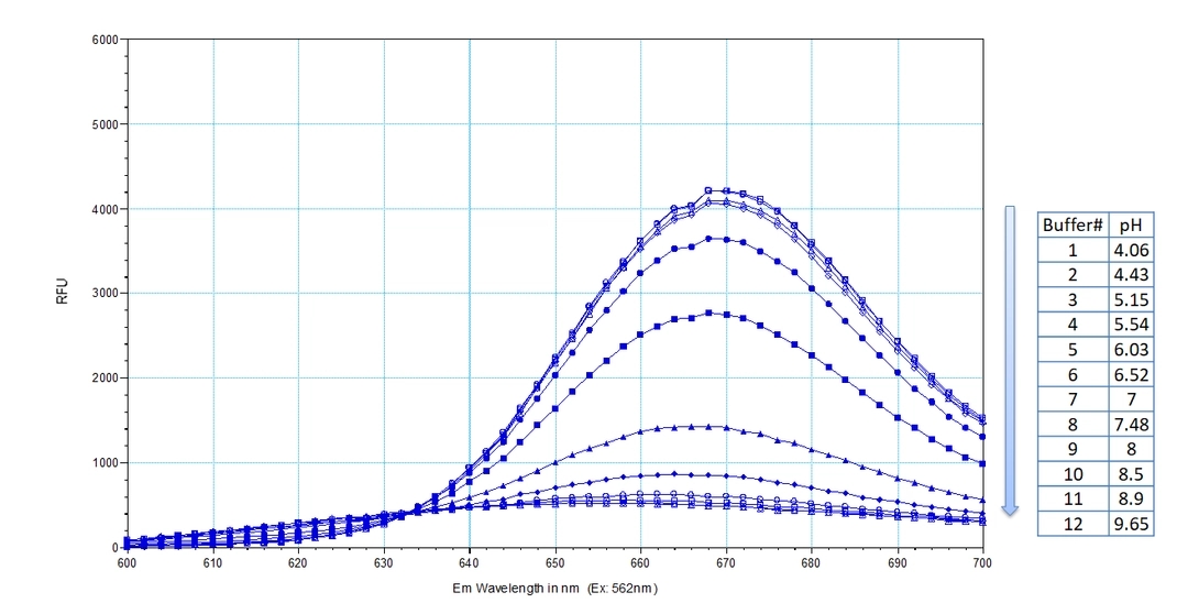
Protonex™ Red 670-Latex Bead Conjugates exhibit unique pH-dependent fluorescence properties. Unlike most existing fluorescent dyes that typically increase in fluorescence at higher pH levels, Protonex™ Red 670-Latex Bead Conjugates become significantly more fluorescent under acidic conditions. This characteristic makes them an ideal tool for studying phagocytosis and its modulation by drugs or environmental factors. The beads display minimal fluorescence outside of cells, eliminating the need for wash steps and enhancing their utility for live-cell imaging. Protonex™ Red 670-Latex Bead Conjugates are particularly effective in highlighting acidic cellular compartments such as phagosomes, lysosomes, and endosomes, where they emit a bright red fluorescence. Additionally, Protonex™ Red 670-Latex Bead Conjugates can be used in combination with green fluorescent dyes like GFP, Fluo-8, calcein, or FITC-labeled antibodies for multiplexed cell functional analysis. Their spectral properties are similar to those of Cy5, allowing for the use of common Cy5 filter sets in assays involving Protonex™ Red 670, further facilitating their integration into existing experimental setups.
Ordering information
Price
Unit size
Catalog Number21222
Quantity
Add to cart
Additional ordering information
Telephone1-800-990-8053
Fax1-800-609-2943
Emailsales@aatbio.com
InternationalSee distributors
Bulk requestInquire
Custom sizeInquire
ShippingStandard overnight for United States, inquire for international
Request quotation
Spectral properties
Excitation (nm)643
Emission (nm)660
Storage, safety and handling
H-phraseH303, H313, H333
Hazard symbolXN
Intended useResearch Use Only (RUO)
R-phraseR20, R21, R22
StorageRefrigerated (2-8 °C); Minimize light exposure
UNSPSC12352200
Platform

Fluorescence microscope

ExcitationCy5 filter set
EmissionCy5 filter set
Recommended plateBlack wall, clear bottom
Example protocol

AT A GLANCE

Chemical and Physical Properties
Solvent:
Water
Solids Content:
1% in PBS
Number of Microspheres per mL:
~4e+10
Ex/Em:
643/660 nm
Mean Diameter:
0.72 µm

SAMPLE EXPERIMENTAL PROTOCOL

Important

The following is a recommended protocol for granulocytes. This protocol only provides a guideline and should be modified
according to your specific experimental conditions. 

Protocol
  1. Prepare cells as desired. For example, prepare the granulocytes at 107 cells/mL with Hanks and 20 mM Hepes buffer (HHBS), and add 100 μL to a polypropylene tube.

    Note: Each cell line should be evaluated on an individual basis to determine the optimal cell density. 

  2. Add 1-10 μL of the Protonex™ Red 670-Latex Bead Conjugate to the tube and incubate with gentle shaking for 30 minutes at 37˚C.

    Note: Each cell line should be evaluated on an individual basis to determine the optimal incubation time. 

  3. Prepare an identical sample that is incubated at 4˚C and label it as a control.

  4. At the end of the 30-minute incubation, stop phagocytosis by adding 2mL of ice-cold HHBS and mix well.

  5. Wash the cells 2 times with cold HBSS. 

  6. Resuspend the cells in 500 μL of cold HBSS, keep the samples at 4˚C, and analyze immediately using a fluorescence microscope equipped with a Cy5 filter set.

    Note: For fluorescence microplate readers, monitor the fluorescence intensity at Ex/Em = 640/680 nm (Cutoff = 660 nm).

Spectrum
Product family
NameExcitation (nm)Emission (nm)
Protonex™ Red 600-Latex Bead Conjugate576597
References
View all 50 references: Citation Explorer
Programmable site-specific delivery of an alkaline phosphatase-activatable prodrug and a mitochondria-targeted cyclopeptide for combination therapy in colon cancer.
Authors: Chen, Huachao and Yao, Yongrong and Zhao, Xing and Tan, Ninghua
Journal: Biomaterials science (2023): 7114-7123
Human Nanoplatelets as Living Vehicles for Tumor-Targeted Endocytosis In Vitro and Imaging In Vivo.
Authors: Dai, Lu and Liu, Yehong and Ding, Shuang and Wei, Xiaowei and Chen, Baoan
Journal: Journal of clinical medicine (2023)
Intracellular trafficking kinetics of nucleic acid escape from lipid nanoparticles via fluorescence imaging.
Authors: M Bailey-Hytholt, Christina and Ulinski, Gregory and Dugas, Julia and Haines, Mohammed and Lazebnik, Mihael and Piepenhagen, Peter and E Zarraga, Isidro and Bandekar, Amey
Journal: Current pharmaceutical biotechnology (2023)
Methods to Quantify the Dynamic Recycling of Plasma Membrane Channels.
Authors: Hodeify, Rawad and Machaca, Khaled
Journal: Bio-protocol (2023): e4800
Macropinoscope: Real-Time Simultaneous Tracking of pH and Cathepsin B Activity in Individual Macropinosomes.
Authors: Hirose, Hisaaki and Nakata, Eiji and Zhang, Zhengxiao and Shibano, Yuya and Maekawa, Masashi and Morii, Takashi and Futaki, Shiroh
Journal: Analytical chemistry (2023): 11410-11419