logo
AAT Bioquest

ReadiPrep™ Nuclear/Cytoplasmic Fractionation Kit

The ReadiPrep™ Nuclear/Cytoplasmic Fractionation Kit offers stepwise isolation of nuclear and cytoplasmic extract from mammalian cells or tissue. The isolated proteins have high concentrations and preserve their biological activities. The nuclear and cytoplasmic extracts from ReadiPrep™ Nuclear/Cytoplasmic Fractionation Kit are compatible with many downstream applications including enzyme activity assays and fluorescent Western blotting etc.
Example protocol

AT A GLANCE

Protocol summary

  1. Rinse cells with PBS
  2. Add 500 µL of Cytosol Extraction Buffer
  3. Centrifuge for 20 seconds
  4. Collect the supernatant (Cytoplasmic extract)
  5. Re-suspend the pellets in 150 µL 1X High Salt Buffer
  6. Centrifuge for 20 minutes
  7. Collect the supernatant (Nuclear Extract)

Important notes
Thaw all the kit components at room temperature before starting the experiment (you may store buffers at 4°C or –20°C), keep the buffers on ice during the experiment.

PREPARATION OF WORKING SOLUTION

1. Cytosol Extraction Buffer (1X):
Add 100 µL of 10X Protease Inhibitors (Component C) and 1 µL of DTT (Component D) into 0.9 mL of Cytosol Extraction Buffer (Component A).

2. Nuclear Extraction Buffer (1X):
Add 100 µL of 10X Protease Inhibitors (Component C) and 1 µL of DTT (Component D) into 0.9 mL of Nuclear Extraction Buffer (Component B).

Note: 0.5 mL 1X Cytosol Extraction Buffer and 150 µL of 1X Nuclear Extraction Buffer is enough for 1 assay, prepare fresh buffer as needed.

For guidelines on cell sample preparation, please visit
https://www.aatbio.com/resources/guides/cell-sample-preparation.html

SAMPLE EXPERIMENTAL PROTOCOL

  1. Rinse the cells 1 time with cold PBS. For cells in suspension, collect the cells by centrifugation.

  2. Add 500 µL of 1X Cytosol Extraction Buffer to cells. For adherent cells, scrape the adherent cells into a 1.5 mL centrifuge tube.

  3. Vortex vigorously to fully re-suspend the cells.

  4. Centrifuge at 16,000 g for 1 - 2 minutes and transfer the supernatant (Cytoplasmic extract) to another clean tube. Keep the tube on ice for downstream applications or store at -80°C.

  5. Re-suspend the pellet in 150 µL 1X Nuclear Extraction Buffer.

  6. Vortex vigorously to fully re-suspend the pellet.

  7. Rotate the tube at 4°C for 30 minutes.

  8. Centrifuge at 16,000g for 20 minutes and transfer the supernatant (Nuclear extract) to another clean tube. Keep the tube on ice for downstream applications or store at -80°C.

Citations

View all 4 citations: Citation Explorer
Circular RNA Hecw1 Regulates the Inflammatory Imbalance in Spinal Cord Injury via miR-3551-3p/LRRTM1 Axis
Authors: Ban, Dexiang and Xiang, Zhenyang and Yu, Peng and Liu, Yang
Journal: Applied Biochemistry and Biotechnology (2022): 5151--5166
Circular RNA circ\_UBAP2 facilitates the progression of osteosarcoma by regulating microRNA miR-637/high-mobility group box (HMGB) 2 axis
Authors: Ma, Weiguo and Zhao, Xin and Gao, Yun and Yao, Xiaobin and Zhang, Junhua and Xu, Qingxia
Journal: Bioengineered (2022): 4411--4427
Circ\_001422 aggravates osteosarcoma progression through targeting miR-497-5p/E2F3 axis
Authors: Li, Xinyu and Zhao, Xin and Li, Jin and Zhang, Xiaozhan
Journal: (2022)
Hsa\_circ\_0088212-mediated miR-520 h/APOA1 axis inhibits osteosarcoma progression
Authors: Liu, Feng and Zhang, Xiangyang and Wu, Fei and Peng, Hao
Journal: Translational Oncology (2021): 101219

References

View all 2 references: Citation Explorer
Studied by Rapid Fractionation of Barley (Hordeum vulgare) Protoplasts
Authors: Gardestrom, P.; Wigge, B., Influence of Photorespiration on ATP/ADP Ratios in the Chloroplasts, Mitochondria, and Cytosol, undefined
Journal: Plant Physiol (1988): 69-76
and mitochondria
Authors: Lilley, R. M.; Stitt, M.; Mader, G.; Heldt, H. W., Rapid fractionation of wheat leaf protoplasts using membrane filtration : the determination of metabolite levels in the chloroplasts, cytosol
Journal: Plant Physiol (1982): 965-70
Page updated on October 4, 2024

Ordering information

Price
Unit size
Catalog Number60000
Quantity
Add to cart

Additional ordering information

Telephone1-800-990-8053
Fax1-800-609-2943
Emailsales@aatbio.com
InternationalSee distributors
Bulk requestInquire
Custom sizeInquire
Technical SupportContact us
Purchase orderSend to sales@aatbio.com
ShippingStandard overnight for United States, inquire for international
Request quotation

Storage, safety and handling

H-phraseH303, H313, H333
Hazard symbolXN
Intended useResearch Use Only (RUO)
R-phraseR20, R21, R22
UNSPSC12352200

Components

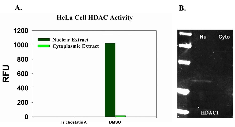
Nuclear and cytoplasmic extract from Hela cells were collected using ReadiPrep&trade; Nucleus/Cytosol Fractionation Kit (AAT Bioquest&reg;, Cat#60000). The protein was quantified by Amplite® Fluorimetric Fluorescamine Protein Quantitation Kit (AAT Bioquest&reg;, Cat#. 11100). <strong>A</strong>. 8 &micro;g of nuclear or cytoplasmic extract was incubated with or without HDAC inhibitor Trichostatin A, and HDAC activity was measured by Amplite® Fluorimetric HDAC Activity Assay Kit (AAT Bioquest&reg;, Cat#. 13601). <strong>B</strong>. 40 ug total protein of nuclear or cytoplasmic extract was used.&nbsp; 2 ug/ml of rabbit anti-HDAC1 antibody was used to probe the nitrocellulose membrane for overnight. 10 ug/ml of iFluorTM 647 goat anti-rabbit IgG (H+L) (AAT Bioquest&reg;, Cat#. 16809) was used. The detection was visualized by UVP MultiSpectral Imaging System (Biolite). M: Marker, Nu: Nuclear extract, Cyto: Cytoplasmic extract.
Nuclear and cytoplasmic extract from Jurkat cells were collected using ReadiPrep&trade; Nucleus/Cytosol Fractionation Kit. The protein was quantified by Amplite® Fluorimetric Fluorescamine Protein Quantitation Kit (AAT Bioquest<sup>&reg;</sup>, Cat#. 11100). 8 &micro;g of nuclear or cytoplasmic extract was incubated with or without HDAC inhibitor Trichostain A, and HDAC activity was measured by Amplite® Fluorimetric HDAC Activity Assay Kit (AAT Bioquest&reg;, Cat#. 13601).
Nuclear and cytoplasmic extract from Hela cells were collected using ReadiPrep&trade; Nucleus/Cytosol Fractionation Kit. The protein was quantified by Amplite® Fluorimetric Fluorescamine Protein Quantitation Kit (AAT Bioquest<sup>&reg;</sup>, Cat#. 11100). 8 &micro;g of nuclear or cytoplasmic extract was incubated with or without HDAC inhibitor Trichostain A, and HDAC activity was measured by Amplite® Fluorimetric HDAC Activity Assay Kit (AAT Bioquest&reg;, Cat#. 13601).
Nuclear and cytoplasmic extract from CHO cells were collected using ReadiPrep&trade; Nucleus/Cytosol Fractionation Kit. The protein was quantified by Amplite® Fluorimetric Fluorescamine Protein Quantitation Kit (AAT Bioquest<sup>&reg;</sup>, Cat#. 11100). 8 &micro;g of nuclear or cytoplasmic extract was incubated with or without HDAC inhibitor Trichostain A, and HDAC activity was measured by Amplite® Fluorimetric HDAC Activity Assay Kit (AAT Bioquest&reg;, Cat#. 13601).
40 ug total protein of nuclear or cytoplasmic extract from Jurkat cells was used.&nbsp; 2 ug/ml of mouse anti-&alpha;-tubulin antibody or rabbit anti-HDAC1 antibody was used to probe the nitrocellulose membrane for overnight. 10 ug/ml of iFluor<sup>TM</sup> 647 goat anti-mouse IgG (H+L) (AAT Bioquest&reg;, Cat#. 16744) or iFluor<sup>TM</sup> 647 goat anti-rabbit IgG (H+L) (AAT Bioquest&reg;, Cat#. 16809) was used. The detection was visualized by UVP MultiSpectral Imaging System (Biolite). M: Marker, Nu: Nuclear extract, Cyto: Cytoplasmic extract.